Post written by Yasuhiro Komori, MD, from the Department of Hepatobiliary and Pancreatic Oncology, National Cancer Center Hospital, Tokyo, and Department of Medicine and Bioregulatory Science, Graduate School of Medical Sciences, Kyushu University, Fukuoka, Japan, Susumu Hijioka, MD, PhD, and Shin Yagi, MD, PhD, from the the Department of Hepatobiliary and Pancreatic Oncology, National Cancer Center Hospital.

During EUS-guided tissue acquisition (EUS-TA), we often encounter a situation during which the needle disappears from the US image during puncture, and the needle tip reappears only after clockwise or counterclockwise torque is applied to the scope. We named this phenomenon the “vanishing needle tract” (VNT) and classified it, according to the direction in which the needle disappears, as clockwise VNT or counterclockwise VNT. In this study, we evaluated the incidence of VNT, its associated factors, and its directional tendency in 1630 lesions that underwent EUS-TA at the National Cancer Center Hospital, Tokyo, Japan.

For safe and accurate EUS-TA, the needle should ideally remain visible on the image throughout the puncture, and VNT can lead to inaccurate targeting of the lesion and inadvertent puncture of vessels. Although VNT is frequently encountered in daily practice, there has been no common term for it, and, to our knowledge, no previous study has examined its incidence or risk factors. We believed that defining VNT and clarifying the situations during which it is likely to occur during EUS-TA would lead to more accurate and safer puncture and to development of new needles.

VNT occurred in 26.6% of lesions, indicating that it is by no means rare. Three-quarters of VNT cases were clockwise, and clockwise VNT predominated in all subgroups examined. On multivariate analysis, puncture via the duodenal bulb (OR, 1.73; 95% CI, 1.28-2.33; P < .001), an increasing number of punctures (OR, 1.45; 95% CI, 1.30-1.62; P < .001), and use of a Franseen needle (OR, 1.70; 95% CI, 1.13-2.58; P = .012) were independent risk factors.
We believe that 2 mechanisms underlie these findings. First, scope maneuverability is restricted in the narrow duodenal bulb. Second, the 3 points of a Franseen needle tip contact the lesion simultaneously, increasing tissue resistance. To our knowledge, this is the first study to define VNT and systematically evaluate its incidence and risk factors.
In clinical practice, when critical structures lie in the clockwise direction relative to the target, endosonographers should be careful about inadvertent puncture caused by VNT. In addition, if the direction of VNT is identified during the first puncture, we suggest applying slight torque in the opposite direction during subsequent punctures. Future validation with different types of echoendoscopes and in multiple centers is warranted, as is the application of these findings to the development of new devices that take VNT into account.
We hope that the new concept of VNT will contribute to the standardization of EUS-TA and serve as a guide for future improvements in needle design.
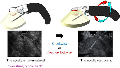
Directional classification of “vanishing needle tract” (VNT). The direction of VNT was classified based on the direction in which the needle disappeared from the EUS image: disappearance in the clockwise direction (clockwise VNT) and disappearance in the counterclockwise direction (counterclockwise VNT).
Read the full article online.
The information presented in Endoscopedia reflects the opinions of the authors and does not represent the position of the American Society for Gastrointestinal Endoscopy (ASGE). ASGE expressly disclaims any warranties or guarantees, expressed or implied, and is not liable for damages of any kind in connection with the material, information, or procedures set forth.

