
Post written by Ariosto Hernández-Lara, MD, MSc, from the Institute of Image-Guided Surgery, Angeles Health System, Strasbourg, France.

In this video, we present proof of concept of indocyanine green (ICG)-guided ERCP in a 30-kg pig that underwent duodenoscopy with a therapeutic colonoscope to reach the pig’s papilla and maintain insufflation while receiving 0.1 mg/kg ICG intravenously. A near-infrared—enabled laparoscope was introduced through an enterotomy in the duodenum to assess the duodenal papilla because no infrared-light endoscopes are available on the market. After identifying the bile duct, we performed a frontal-view cannulation of the bile duct with a precut knife (Fusion Needle Knife; Cook Medical, Bloomington, Ind, USA).
Post-ERCP adverse events such as bleeding and pancreatitis can be avoided with higher cannulation rates. Small papillas (type 2) and those with difficult anatomy, such as diverticula, can be challenging to cannulate. Technologies such as narrow-band imaging are available in endoscopy to improve detection and characterization of areas of interest in the digestive tract.
ICG is used in hepatobiliary surgery to identify the common bile duct and to avoid adverse events. We believe ICG-enhanced near-infrared imaging could help endoscopists identify the papilla orifice in scenarios where the anatomy is challenging and find the common bile duct once it is exposed when advanced cannulation techniques such as precut and fistulotomy are performed. To our knowledge, this is the first time that images of the papilla are documented under ICG near-infrared technology from the inside of the duodenum.
We believe that enhanced near-infrared technology and ICG applied to endoscopy could expand its applications. Other narrow-band imaging technologies are already available in the scopes we use, and companies specialized in endoscopy could invest their efforts to incorporate this technology, as some are already applying it in other fields of surgery.
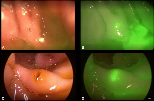
ICG/NIR localization overtime. A, White-light view of the papillary region approximately 5 minutes after IV ICG. B, NIR view approximately 5 minutes after IV ICG showing ICG-enhanced bile outflow at the orifice. C, White-light view 1 hour after IV ICG. D, NIR view 1 hour after IV ICG showing increased background fluorescence with the papillary region still identifiable. ICG, Indocyanine green; IV, intravenous; NIR, near-infrared.
Read the full article online.
The information presented in Endoscopedia reflects the opinions of the authors and does not represent the position of the American Society for Gastrointestinal Endoscopy (ASGE). ASGE expressly disclaims any warranties or guarantees, expressed or implied, and is not liable for damages of any kind in connection with the material, information, or procedures set forth.

