
Post written by Yuki Amioka, MD, and Hidenori Tanaka, MD, PhD, from the Department of Gastroenterology, Hiroshima University Hospital, Hiroshima, Japan.

Laterally spreading tumors (LSTs) of the nongranular type are classified into flat-elevated (FE) and pseudodepressed (PD) subtypes, which have different malignant potential. Although magnifying endoscopy is widely used to predict histology and invasion depth in colorectal neoplasia, its clinical value for therapeutic decision-making with LSTs of the nongranular type remains unclear. We evaluated 282 consecutive LSTs of the nongranular type, including 250 FE and 32 PD lesions, to determine whether magnifying narrow-band imaging and magnifying chromoendoscopy provide clinically meaningful information for selecting an appropriate treatment strategy.

PD lesions have a particularly high risk of submucosal invasion and may exhibit multifocal invasion. Accurate assessment is therefore important for selecting between endoscopic and surgical treatment and achieving reliable histopathological evaluation. However, the relationship between magnifying endoscopic findings and histopathology in LSTs of the nongranular type, particularly PD lesions, has not been sufficiently investigated. We therefore sought to clarify whether detailed magnifying observation actually provides information that changes therapeutic decision-making.
PD lesions showed significantly higher rates of T1 carcinoma and T1b carcinoma than FE lesions (34.4% vs 2.0% and 9.4% vs 0%, respectively). Nearly all PD lesions were classified as Japan Narrow-band Imaging Expert Team type 2B, despite histological findings ranging from low-grade dysplasia to T1b carcinoma. Furthermore, all 3 PD lesions with T1b carcinoma showed homogeneous magnifying endoscopic findings but multifocal submucosal invasion, indicating that magnifying endoscopy could not precisely identify the depth or location of invasion.
These findings suggest that, for PD lesions, magnifying endoscopy provides limited additional information for therapeutic decision-making. When technically feasible, en bloc endoscopic resection, particularly by ESD, may therefore be an appropriate default strategy regardless of magnifying endoscopic findings. Further multicenter studies are needed to validate this treatment strategy in larger and more diverse populations.
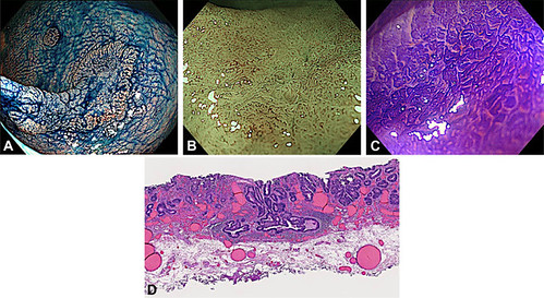
Representative case of submucosal invasion in LST-NG (pseudodepressed type [PD]). A, A 35-mm LST-NG (PD subtype) located in the rectum. B, Magnifying endoscopic findings of the pseudodepressed area showing a homogeneous JNET type 2B by narrow-band imaging. C, Crystal violet staining of the pseudodepressed area showing a homogeneous VI low-grade pit pattern. D, Low-power view of the pathologic specimen stained with hematoxylin and eosin. The submucosal invasion depth was measured at 1700 μm. JNET, Japan Narrow-band Imaging Expert Team; LST-NG, laterally spreading tumor nongranular type.
Read the full article online.
The information presented in Endoscopedia reflects the opinions of the authors and does not represent the position of the American Society for Gastrointestinal Endoscopy (ASGE). ASGE expressly disclaims any warranties or guarantees, expressed or implied, and is not liable for damages of any kind in connection with the material, information, or procedures set forth.

