
Post written by Babak Mirminachi, MD, MPH, and Sultan Mahmood, MD, from the University of Pittsburgh Medical Center, Pittsburgh, Pennsylvania, USA.

Our video shows endoscopic resection of a distal rectal GI stromal tumor located 1.5 cm proximal to the dentate line using endoscopic intermuscular dissection. EUS initially suggested a submucosal origin, but during endoscopic submucosal dissection, the lesion was found to arise from the muscularis propria.
We therefore entered the plane between the circular and longitudinal muscle layers and achieved en bloc, R0 resection with an intact capsule while preserving the anorectal anatomy. The defect was closed with an endoscopic suturing device. The procedure was completed without adverse events, and the patient remained recurrence-free at 14-month follow-up.

This article demonstrates the feasibility and effectiveness of intermuscular dissection for complete, organ-preserving resection in a technically challenging, very distal location, and it illustrates a real-time intraoperative pivot when the true layer of origin differed from the preprocedural assessment.
Other endoscopists can learn from this experience that:
- Endoscopic intermuscular dissection is a feasible, organ-preserving option for selected distal rectal lesions that do not extend beyond the circular layer of the muscularis propria.
- Conventional techniques have limits: EMR yields lower en bloc and R0 rates, and the full-thickness resection device’s bulky cap, limited tissue capture, and pain near the dentate line restrict its use in very distal lesions.
- Because the intermuscular space is far narrower than the submucosal plane, special care is needed to open the plane for safe, precise dissection as highlighted in the video.
- EUS may not reliably identify the true layer of origin of a subepithelial lesion, and the plane of dissection may need to be adjusted based on the intraprocedural findings.
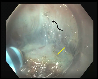
Wall layers with exposed intermuscular space illustrated. The curved black arrow indicates the outer longitudinal layer, and the yellow arrow indicates the tumor capsule involving the circular layer.
Read the full article online.
The information presented in Endoscopedia reflects the opinions of the authors and does not represent the position of the American Society for Gastrointestinal Endoscopy (ASGE). ASGE expressly disclaims any warranties or guarantees, expressed or implied, and is not liable for damages of any kind in connection with the material, information, or procedures set forth.

