
GIE Associate Editor Seiichiro Abe, MD, PhD, FASGE, FJGES, highlights this article from the April issue: “Computer-aided characterization of early cancer in Barrett’s esophagus on i-scan magnification imaging: a multicenter international study” by Mohamed Hussein, MRCP, et al.

Magnifying virtual chromoendoscopy is helpful in differentiating between dysplastic and nondysplastic lesions in patients with Barrett’s esophagus (BE), and some classification systems are available.
However, assessment on magnification images can be difficult in real clinical practice, especially for nonexpert endoscopists.
The authors of this study developed a novel computer-aided diagnosis (CAD) system that could characterize and diagnose BE dysplasia on optical enhancement/i-scan3 magnification endoscopic imaging. The CAD can characterize the lesions in BE with high accuracy and speed on high-quality magnification images and sequences of video frames.
The artificial intelligence—assisted magnifying endoscopy allows for accurate diagnosis and optimal treatment decisions and follow-up in patients with BE. This CAD system can move it forward to real-time automated diagnosis in clinical practice. The key point should be how endoscopists can receive sufficient training and provide high-quality magnifying endoscopy.
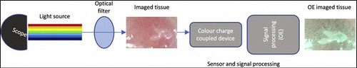
Before and after application of the i-scan optical enhancement (OE) technology during assessment of an area of Barrett’s esophagus on magnification imaging. Image courtesy of Everson et al, 2019.
Read the full article online.
The information presented in Endoscopedia reflects the opinions of the authors and does not represent the position of the American Society for Gastrointestinal Endoscopy (ASGE). ASGE expressly disclaims any warranties or guarantees, expressed or implied, and is not liable for damages of any kind in connection with the material, information, or procedures set forth.
