
Post written by Bing Hu, MD, from the Department of Gastroenterology, West China Hospital, Sichuan University, Chengdu, Sichuan, China.

The focus of this study was to train an artificial intelligence (AI) system that could detect and delineate the extent of superficial esophageal squamous cell carcinoma (ESCC) and precancerous lesions under nonmagnified narrow-band imaging (NBI) and to validate the diagnostic performance of the AI system from multiple perspectives.
Early detection and treatment are crucial to improving the prognosis of patients with ESCC. Effective endoscopic biopsy of lesions is the key to ensuring no missed lesions or misdiagnosis of lesions. Accurate delineation of the extent of the lesions is essential for biopsy.
To date, the application of AI in delineating the extent of superficial ESCC and precancerous lesions under NBI has rarely been investigated.
A deep convolutional neural network model called You Only Look At CoefficienTs (YOLACT) could accurately detect superficial ESCC and precancerous lesions and delineate the extent of lesions under nonmagnified NBI. Real-world studies are needed to further evaluate the auxiliary role of the AI system for endoscopists with varying endoscopic experience.
To our knowledge, this is the first report evaluating the ability of the AI system to accurately delineate the extent of superficial ESCC and precancerous lesions under nonmagnified NBI. Unlike other previously reported AI systems that require a separate monitor, the proposed AI system was directly connected to the endoscopic monitor commonly used by endoscopists without changing their operating habits, which was more suitable for clinical use.
In conclusion, we hope that the AI system can promote the early diagnosis of ESCC and guide the rational establishment of treatment plans in the future.
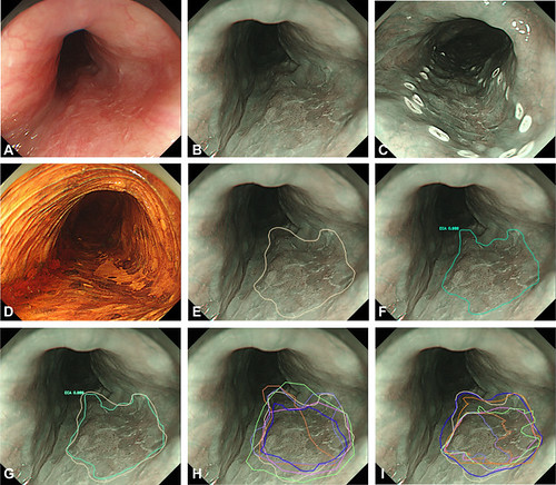
A representative cancerous lesion (high-grade intraepithelial neoplasia) detected and delineated by the artificial intelligence (AI) system and endoscopists. A, White-light imaging. B, Narrow-band imaging. C, Intraoperative electrocautery markers. D, Iodine staining. E, The extent of the lesion was manually delineated by an expert and used as the criterion standard. F, The AI system detected and delineated the lesion by indicating it with a polygon. G, Comparison of the extent of the lesion delineated by the AI system and the criterion standard. H, Comparison of the extent of the lesion delineated by 5 senior endoscopists and the criterion standard. I, Comparison of the extent of the lesion delineated by 6 junior endoscopists and the criterion standard.
Read the full article online.
The information presented in Endoscopedia reflects the opinions of the authors and does not represent the position of the American Society for Gastrointestinal Endoscopy (ASGE). ASGE expressly disclaims any warranties or guarantees, expressed or implied, and is not liable for damages of any kind in connection with the material, information, or procedures set forth.
