
Ming-Yan Cai, MD, from the Endoscopy Center and Endoscopy Research Institute, Zhongshan Hospital, Fudan University, Shanghai, China, presents this video case. 
During a regular EGD exam, using a magnifying scope with narrow-band imaging (NBI) function, we observed a very interesting pattern of Ancylostoma duodenale. A male worm was coiled around a female one and the head of female worm was attached to the intestinal wall. It was very interesting to view the biological behavior of Ancylostoma duodenale in the duodunem. A direct diagnosis of hookworm infection was confidently made by viewing this pattern.
High-resolution magnifying endoscopy with water-immersion technique can provide an ideal visualization of duodenal mucosa.
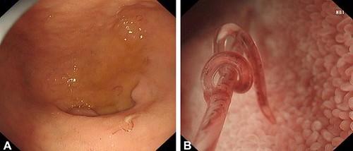
Figure 1. A, EGD view showing several red short worms in the duodenal bulb. B, Narrow-band imaging and magnification showing a male worm coiled around a female worm.
Find more VideoGIE cases online.
The information presented in Endoscopedia reflects the opinions of the authors and does not represent the position of the American Society for Gastrointestinal Endoscopy (ASGE). ASGE expressly disclaims any warranties or guarantees, expressed or implied, and is not liable for damages of any kind in connection with the material, information, or procedures set forth.
