
Post written by Shamira Ghouse, MRCP, from the Gastroenterology Department, East and North Hertfordshire NHS Teaching Trust, Lister Hospital, Stevenage, England.

Our article describes a 42-year-old man with no relevant medical history who was referred on the urgent suspected cancer pathway with new intermittent abdominal pain and an altered bowel habit. Colonoscopy showed a subepithelial polypoidal lesion exceeding 50 mm in the distal descending colon, causing intussusception. Cross-sectional imaging confirmed a 60-mm mass with appearances typical of a lipoma and intussusception spanning 13 cm of bowel.
Biopsy findings revealed ulceration and mucosal prolapse, with no evidence of malignancy. Our multidisciplinary meeting recommended a left hemicolectomy, but the patient declined surgery and was referred for a trial of endoscopic deroofing.
Forty-eight hours before the planned procedure and before any bowel preparation, the patient had a short episode of severe pain and then passed a large pale mass per the rectum. This was 6 weeks after the index colonoscopy. Follow-up colonoscopy showed an ulcerated area of breached mucosa at the prior lesion site, no residual lipoma, and complete reduction of the intussusception. Histology of the expelled specimen confirmed a submucosal lipoma.
This outcome changed the plan from major surgery or endoscopic resection to observation, and we felt it was worth sharing because, to our knowledge, spontaneous expulsion of a lipoma this large is exceedingly rare.
Our patient was asymptomatic at clinic review 4 months later, so no further endoscopic or radiological follow-up was deemed necessary. Further reports will help clarify the mechanism and which patients can safely be observed.
The mechanism remains uncertain. The few documented cases suggest expulsion is more likely in lipomas with long pedunculated stalks, which may predispose to torsion, ischemia, ulceration, and eventual separation. Some reported cases were associated with bowel preparation for colonoscopy, but in our patient, expulsion happened before any preparation. Management in the literature ranges from conservative observation, when expulsion is complete and symptoms resolve, to endoscopic or surgical intervention when adverse events arise or diagnostic uncertainty persists.
Adult intussusception caused by lipomas is uncommon, and spontaneous expulsion of large colonic lipomas is rarer still. It was first described by Backenstoe et al1,2 in 1940, and since then, only a small number of cases have been reported, almost exclusively as isolated case reports or limited case series. Published reports suggest that expulsion is typically preceded by abdominal pain and bleeding, which, as in our patient, often resolve once the lesion detaches.
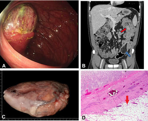
A, Endoscopic image from the index colonoscopy demonstrating the subepithelial lesion found. B, Coronal cross-sectional imaging showing the mass (blue arrow) and the associated segment of intussuscepted bowel (red arrow). C, The mass that was spontaneously passed by the patient. D, Histologic section stained with hematoxylin and eosin, original magnification ×10, showing benign mucosa with submucosal distribution of the lipoma (red arrow).
Visit iGIE’s Facebook, X/Twitter, LinkedIn, Instagram, and YouTube accounts for more content from the ASGE peer-reviewed journal that launched in December 2022 and has been PubMed indexed.
Read the full article online.
The information presented in Endoscopedia reflects the opinions of the authors and does not represent the position of the American Society for Gastrointestinal Endoscopy (ASGE). ASGE expressly disclaims any warranties or guarantees, expressed or implied, and is not liable for damages of any kind in connection with the material, information, or procedures set forth.
- Hamila F, Elghali MA, Bouriga R, et al. Partial spontaneous anal expulsion of the right colon lipoma: an exceptional diagnostic circumstance. Int J Surg Case Rep 2017;41:414-6. ↩︎
- Kouritas VK, Baloyiannis I, Koukoulis G, et al. Spontaneous expulsion from rectum: a rare presentation of intestinal lipomas. World J Emerg Surg 2011;6:19. ↩︎

