Post written by Abdulrahman Qatomah, MBBS, from the Division of Gastroenterology, Hepatology and Endoscopy, Brigham and Women’s Hospital, Harvard Medical School, Boston, Massachusetts, USA, and Department of Medicine, King Faisal Specialist Hospital and Research Center, Jeddah, Saudi Arabia, and Marvin Ryou, MD, from the Division of Gastroenterology, Hepatology and Endoscopy, Brigham and Women’s Hospital, Harvard Medical School.

This video is a step-by-step guide to performing EUS-guided variceal therapy (EUS-VT) for cardiofundal varices, from room set-up to postprocedural surveillance. We cover team and room layout, the equipment tray, variceal measurement and how it drives needle and coil selection, coil loading, preparation of the adjunct hemostatic agent, water-assisted visualization, and feeder vessel targeting. We then walk through the pitfalls we encounter most often, including needle-coil mismatch, coil migration, and eroded coils, and show how to prevent or manage each.
EUS-VT achieves higher obliteration rates with fewer sessions than direct glue injection, but those results depend on technique and team preparation. Much of what makes the procedure work, such as where everyone stands, how the coil is loaded, and how the needle is matched to the coil, is learned at the bedside and rarely written down. We wanted to bring the entire workflow together, so that teams starting EUS-VT have a practical, complete guide.
Measure the varix first and let the diameter guide the equipment. A 0.035-inch coil through a 19-gauge needle suits varices larger than 5 mm, and a 0.018-inch coil through a 22-gauge needle suits smaller ones. Pairing a 0.018-inch coil with a 19-gauge needle is a common cause of failed deployment because the stylet cannot reliably advance the coil. Choose a coil about 30% larger than the varix and anchor the first 1 across a septum with the sewing technique. Preload the first coil, and when advancing the stylet, leave 25 to 30 cm outside the needle to avoid premature deployment.
For the gelatin sponge, dilute to a total of 10 mL saline for a 19-gauge needle or 20 mL for a 22-gauge needle. Instilling 100 to 200 mL of water helps trace the varix to its feeder vessel, and Doppler should confirm reduced flow before injecting glue or gelatin sponge.
Safety starts before the procedure, with cross-sectional imaging to define shunt anatomy. Eroded coils can be cut with endoscopic scissors and removed. Our protocol is repeat EUS every 4 weeks until obliteration, then at 12 months.
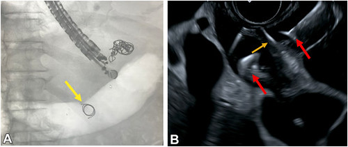
A, Fluoroscopic image showing a migrated coil during EUS-guided variceal therapy (yellow arrow). B, EUS image of the sewing technique with proximal and distal coils (red arrows) deployed across the intervariceal septum (orange arrow).
Read the full article online.
The information presented in Endoscopedia reflects the opinions of the authors and does not represent the position of the American Society for Gastrointestinal Endoscopy (ASGE). ASGE expressly disclaims any warranties or guarantees, expressed or implied, and is not liable for damages of any kind in connection with the material, information, or procedures set forth.

