
Post written by Noel Ravindranayagam, MBBS (Hons), from the Department of Gastroenterology, Surgical Treatment and Rehabilitation Service, Herston, and Rajit Gilhotra, MBBS (Hons), AFRACMA, FRACP, from the Department of Gastroenterology, Royal Brisbane and Women’s Hospital, Brisbane, Australia.

This case report illustrates the rare, unusual finding of hemostatic spray (in this case, Hemospray; Cook Medical, Bloomington, Ind, USA) visualized on computerized tomography imaging after endoscopic application in the management of upper gastrointestinal bleeding.
Hemostatic spray is an established tool that is increasingly used for the endoscopic control of nonvariceal upper gastrointestinal bleeding by binding to applied surfaces to form a mechanical barrier, arresting bleeding. Hemostatic spray is not thought to be radio-opaque because of its chemical components not being radio-opaque and thus should not appear on radiological imaging postapplication. However, if deployed into a narrow-enough lumen and mixed with a high-enough volume of blood, it may appear radio-opaque, as was the situation in this case report.

To our knowledge, this represents the first reported case of endoscopically applied hemostatic spray visualized on cross-sectional imaging. Clinicians should remain mindful of this phenomenon, especially when imaging patients following powder application in narrow, blood-filled lumens. Distinguishing this material from active bleeding is critical; although both appear as hyperdense material within the lumen, the hemostatic powder is uniquely characterized by its high density on noncontrast phases and its stability across subsequent imaging acquisitions. In this case, repeat upper endoscopy confirmed control of bleeding posthemostatic spray application with no recurrence clinically.
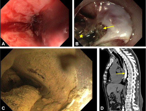
A, Endoscopic view of the distal esophagus, demonstrating Los Angeles grade D esophagitis with active bleeding. B, Endoscopic view of the culprit vessel in the distal esophagus (arrow) after application of focal coagulation graspers (star). C, Endoscopic view of the distal esophagus after application of Hemospray (Cook Medical, Bloomington, Ind, USA). D, Sagittal noncontrast-phase computed tomography of the chest, with the radio-opaque Hemospray within the esophagus (arrow).
Visit iGIE’s Facebook, X/Twitter, LinkedIn, Instagram, and YouTube accounts for more content from the ASGE peer-reviewed journal that launched in December 2022 and has been PubMed indexed.
Read the full article online.
The information presented in Endoscopedia reflects the opinions of the authors and does not represent the position of the American Society for Gastrointestinal Endoscopy (ASGE). ASGE expressly disclaims any warranties or guarantees, expressed or implied, and is not liable for damages of any kind in connection with the material, information, or procedures set forth.

