
Post written by Shyam Thakkar, MD, and Ayowumi A. Adekolu, MD, from the Department of Gastroenterology & Hepatology, West Virginia University, Morgantown, West Virginia, USA.

We present 2 cases involving patients with undifferentiated pancreatic lesions (pancreatic head in case 1 and pancreatic uncinate process in case 2) concerning for malignancy who underwent EUS-guided sampling. In case 2, the patient received prophylactic ciprofloxacin before and for 5 days after the procedure, but not in case 1, given the patient’s history of multiple antibiotic allergies.

Both cases had benign findings on histology. However, after EUS-guided sampling, the patients developed sepsis and underwent successful EUS-guided transmural drainage because of concern for infected lesions, and they were treated with antibiotics. Histologic analysis revealed keratinizing squamous epithelium with underlying lymphocytes, diagnostic of lymphoepithelial cysts (LECs).
We felt it was important to share the video case because, when evaluating pancreatic cystic lesions (PCLs), delineation of their malignant potential is essential for management. An LEC is a PCL type that can resemble a pancreatic neoplasm radiologically and can pose a diagnostic challenge. EUS-FNA and fine-needle biopsy (FNB) are diagnostic tools for LECs. The American Society for Gastrointestinal Endoscopy recommends prophylactic antibiotics for EUS-FNA evaluation of cystic pancreatic neoplasms. Data have shown that the infection rate after EUS-FNA of PCLs is low and have not supported the routine use of prophylactic antibiotics.
However, in our 2 cases, systemic infection occurred after instrumentation of LECs despite antibiotic prophylaxis in 1 of the cases. Furthermore, to our knowledge, in the only other reported case1 of an infected LEC in the literature, infection occurred spontaneously. This raises the concern that LECs may carry a higher risk of infection.
Several key insights can be ascertained from our case:
- LECs pose a diagnostic challenge because they can resemble pancreatic neoplasms radiologically and often require EUS-FNA or FNB for diagnosis.
- Although data are conflicting on the role of prophylactic antibiotics after EUS sampling of pancreatic cysts, given low infection rate, we hypothesize that LECs may carry a higher risk of infection. Therefore, prophylactic antibiotics should be considered with close observation and follow-up, given the risk of sepsis observed in our series despite prophylactic antibiotic use.
- Infected LECs can be managed via EUS-guided drainage and debridement with favorable outcomes.
In patients who undergo EUS-FNA or FNB of a PCL and are found to have an LEC or in whom there is concern for an LEC, close follow-up and care should be considered, given the concern for higher infection risk.
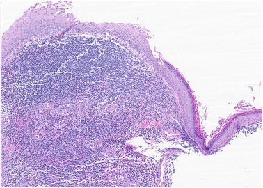
Low-power hematoxylin and eosin–stained section demonstrating a well-circumscribed cyst lined by mature stratified squamous epithelium with subjacent dense lymphoid tissue diagnostic of lymphoepithelial cyst. Original magnification ×100; scale bar = 200 μm.
Read the full article online.
The information presented in Endoscopedia reflects the opinions of the authors and does not represent the position of the American Society for Gastrointestinal Endoscopy (ASGE). ASGE expressly disclaims any warranties or guarantees, expressed or implied, and is not liable for damages of any kind in connection with the material, information, or procedures set forth.
- Chowdhry M, Bilal M, Shah H, et al. Large lymphoepithelial cyst of pancreas: a rare entity managed using lumen-apposing metal stent-case report and review of the literature. Clin J Gastroenterol 2019;12:609-14. ↩︎

