
Post written by Matteo Tacelli, MD, PhD, from the Pancreato-Biliary Endoscopy and Endosonography Division, Pancreas Translational and Clinical Research Center, IRCCS San Raffaele Scientific Institute, Milan, Italy.

Can we move contrast-enhanced endoscopic ultrasound (CE-EUS) from a subjective visual impression to an objective and reproducible diagnostic tool? Our study explored whether quantitative analysis of CE-EUS using dedicated perfusion software (VueBox software; Bracco Suisse SA, Geneva, Switzerland) can reliably differentiate pancreatic ductal adenocarcinomas from pancreatic neuroendocrine neoplasms. By transforming contrast enhancement into measurable perfusion parameters, we aimed to reduce operator dependency and improve the diagnostic value of CE-EUS.
Despite the widespread use of CE-EUS, its interpretation still largely depends on the endosonographer’s subjective assessment. Interestingly, previous studies have demonstrated that even experienced experts frequently disagree when interpreting contrast enhancement patterns. We believed it was time to investigate whether objective quantitative analysis could overcome this limitation and provide standardized biomarkers that better reflect the biological differences between pancreatic tumors.
Our prospective study showed that several quantitative perfusion parameters differed significantly between pancreatic ductal adenocarcinomas and pancreatic neuroendocrine neoplasms, with the combination of wash-in perfusion index and wash-in rate achieving an excellent diagnostic performance (area under the curve 0.96). These findings suggest that quantitative CE-EUS captures intrinsic tumor vascularity while minimizing subjective interpretation.
More importantly, this study represents an initial step toward standardization of quantitative CE-EUS. Future multicenter studies should validate these biomarkers across different centers and ultrasound platforms, define clinically applicable cutoff values, and explore whether quantitative perfusion analysis can also predict tumor aggressiveness, treatment response, and prognosis, bringing CE-EUS closer to precision medicine.
Quantitative CE-EUS has the potential to transform how we interpret pancreatic lesions—not by replacing the endosonographer, but by complementing clinical expertise with objective, reproducible imaging biomarkers. We hope this work stimulates further multicenter collaborations toward standardization of quantitative endoscopic ultrasound imaging.
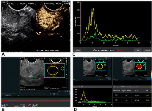
Workflow of qCE-EUS analysis using VueBox (Bracco Suisse SA, Geneva, Switzerland). A, Left: B-mode endoscopic ultrasound image shows a round hypoechoic solid pancreatic mass. Right: synchronized CE-EUS frame 12 seconds after contrast administration (arterial phase) demonstrating hyperenhancement of the lesion compared with the surrounding parenchyma. B, ROI placement on the CE-EUS harmonic image: the tumor ROI (yellow) completely encompasses the pNEN, whereas the reference ROI (green) is placed in visually normal pancreatic parenchyma. ROIs are size-matched and positioned to avoid vessels and ducts. C, TICs for the 2 ROIs. Y-axis: echo-power/intensity (a.u.). X-axis: time (s). The vertical red cursor indicates the user-selected contrast-arrival time, which is used to define the analysis windows. D, Final VueBox output with motion-corrected and normalized TICs illustrating the PE parameter. The yellow curve refers to the tumor ROI and the green curve to the reference parenchyma. The table summarizes the mean PE for each ROI. a.u., Arbitrary unit; CE-EUS, contrast-enhanced endoscopic ultrasound; DICOM, Digital Imaging and Communications in Medicine; PE, peak enhancement; pNEN, pancreatic neuroendocrine neoplasm; qCE-EUS, quantitative contrast-enhanced endoscopic ultrasound; ROI, region of interest; TIC, time-intensity curve.
Visit iGIE’s Facebook, X/Twitter, LinkedIn, Instagram, and YouTube accounts for more content from the ASGE peer-reviewed journal that launched in December 2022 and has been PubMed indexed.
Read the full article online.
The information presented in Endoscopedia reflects the opinions of the authors and does not represent the position of the American Society for Gastrointestinal Endoscopy (ASGE). ASGE expressly disclaims any warranties or guarantees, expressed or implied, and is not liable for damages of any kind in connection with the material, information, or procedures set forth.

