
Post written by Samer Al-Dury, MD, PhD, from Muscat Endoscopy Academy, Endoscopy Unit, Division of Gastroenterology and Hepatology, Medical City for Military and Security Services, Muscat, Oman.

This video presents a low-cost simulator designed specifically for hands-on training in both GI bleeding control and perforation management. The platform can use ex vivo animal tissue or, where available, 3-dimensional—printed tissue. Multiple bleeding sites and perforation defects can be created at different locations and angles, allowing scenarios to be arranged in tiers of increasing technical difficulty. Trainees can therefore progress from straightforward cases to more challenging situations requiring precise targeting, hemostasis, defect closure, and suturing.
Commercial endoscopy simulators are available and provide valuable training opportunities, but cost and access can limit their regular use. In many settings, such training is concentrated in larger centers or encountered mainly during courses, conferences, and workshops. We wanted to demonstrate that meaningful simulation also can be created locally using inexpensive, readily available materials without requiring complex engineering expertise. This makes repeated practice within the trainee’s own unit much more achievable.
A key lesson is that simulation does not need to reproduce 1 standardized model. What matters is creating a safe environment where trainees can repeatedly practice management of bleeding and perforation, receive immediate feedback, and progress through increasingly difficult scenarios. By varying the site, orientation, and number of bleeding sources or defects, the same platform can support both early skills acquisition and more advanced technical refinement.
Importantly, it also allows theoretical or virtual learning to be followed immediately by hands-on practice. Concepts learned from lectures, videos, or online teaching can be translated directly into procedural experience, helping consolidate knowledge and increasing familiarity with difficult situations before they occur in patients. The model is intended to complement, rather than replace, supervised clinical training.
This project reflects a broader aim of the Muscat Endoscopy Academy: to make high-quality endoscopy training more accessible, practical, and reproducible. We hope it encourages other centers to adapt simulation to their own resources and integrate repeated hands-on practice into everyday endoscopy education. A next step will be to evaluate how structured use of the model affects technical performance and trainee confidence across different levels of experience.
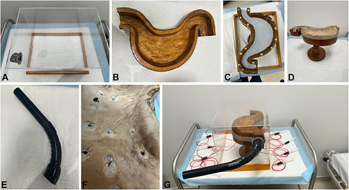
Equipment and mounting of a Muscat Endoscopy Academy Gastric Simulator. A, Acrylic enclosure. B, Carved wooden stomach mold. C, Silicone-lined mold. D, Ex vivo stomach mounted on the mold. E, Detachable entry tube. F, Artificially created tissue defects. G, Fully assembled simulator.
Read the full article online.
The information presented in Endoscopedia reflects the opinions of the authors and does not represent the position of the American Society for Gastrointestinal Endoscopy (ASGE). ASGE expressly disclaims any warranties or guarantees, expressed or implied, and is not liable for damages of any kind in connection with the material, information, or procedures set forth.

