Post written by Rizwan Ahamed, MD, DM, Sasidharan Rajesh, MD, PDCC, and Cyriac Abby Philips, DM, from the Division of Gastroenterology and Division of Hepatobiliary Interventional Radiology, Center of Excellence in GI Sciences, Rajagiri Hospital, Kochi, India.

A 41-year-old man with alcohol-related Child-Pugh B cirrhosis was referred to us after failed endoscopic glue injection for bleeding fundal varices. CT showed a large gastrorenal shunt fed by 2 afferent veins, the left gastric and posterior gastric veins, that drained into a single efferent channel entering the left renal vein.
Instead of targeting the multiple feeders, we occluded the single efferent under EUS guidance. Three fibered coils, oversized by 20%, were deployed through a 19-gauge FNA needle, followed by lipiodol glue injection. This achieved complete obliteration of the variceal complex. The patient remained asymptomatic without rebleeding at 1 year.
Conventional EUS-guided therapy targets the varix itself or its afferent feeders. When several feeders are present, this may leave residual flow and incomplete obliteration. By tracing and closing the single efferent, we reproduced the end point of interventional radiology–guided balloon–occluded retrograde transvenous obliteration through the endosonographic route.

To our knowledge, this efferent-directed EUS technique, which we term EUS-guided coil–assisted retrograde transgastric occlusion (e-CARTO), has not been described before. It offers a lower-cost option where interventional radiology expertise or funding is limited.
The main lesson is conceptual. When a gastric varix has multiple afferents but a single outflow, occluding the efferent produces complete stasis where feeder-directed therapy can fail. Useful technical points include oversizing coils by about 20% to prevent nontarget embolization, mixing lipiodol with glue in a 1:1 ratio for fluoroscopic visibility, and selecting the efferent segment just distal to the varix to reduce embolic risk. As with balloon–occluded retrograde transvenous obliteration, shunt closure can promote new esophageal varices, so endoscopic surveillance remains essential.
The treatment plan followed a multidisciplinary discussion, and combined fluoroscopic and EUS guidance was central to safe coil and glue delivery. Larger series are needed to confirm durability and refine patient selection.
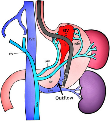
Schematic diagram showing the gastric variceal complex anatomy with 2 afferent feeders and a single efferent outflow. GV, Gastric varix; IVC, inferior vena cava; LGV, left gastric vein; LRV, left renal vein; PGV, posterior gastric vein; PV, portal vein; SMV, superior mesenteric vein; SV, splenic vein.
Read the full article online.
The information presented in Endoscopedia reflects the opinions of the authors and does not represent the position of the American Society for Gastrointestinal Endoscopy (ASGE). ASGE expressly disclaims any warranties or guarantees, expressed or implied, and is not liable for damages of any kind in connection with the material, information, or procedures set forth.
 endoscopedia.com