Post written by Connor John Rodriguez, MD, and Robert Jay Sealock, MD, from Baylor College of Medicine, Ben Taub Hospital, Houston, and University of North Texas Health, Fort Worth, Texas, USA.
We present the case of a 52-year-old man who developed a chronic sigmoid cutaneous fistula following abdominal operations after a severe motor vehicle collision.
Despite fecal diversion, percutaneous drainage, and prior endoscopic closure attempts with helical tack—based (Boston Scientific, Marlborough, Mass, USA) and full-thickness suturing, the fistula persisted.
We therefore pursued endoscopic vacuum therapy (EVT) using a custom open-pore sponge connected to a nasogastric tube and continuous negative pressure. Serial endoscopic exchanges resulted in progressive granulation tissue formation and contraction of the fistula cavity, ultimately achieving complete closure.

EVT is increasingly recognized as an effective treatment for GI defects, but its use in refractory lower GI fistulas remains less commonly reported. We felt this case was important because it demonstrates how EVT can provide a minimally invasive, organ-preserving treatment option even after conventional endoscopic closure techniques have failed. We chose to present this case in video format to allow viewers to directly visualize the technique and progressive changes in the fistula cavity during therapy, which are difficult to convey through text and still images alone.
This case highlights that successful management of a complex fistula does not always require immediate mechanical closure of the defect. EVT instead promotes healing by controlling the local wound environment and encouraging granulation and progressive cavity contraction. Careful sponge positioning and serial endoscopic reassessment are important components of treatment. This case also shows the value of adapting EVT technique as the cavity decreases in size and reinforces the role of EVT as an effective therapy for selected refractory lower GI defects.
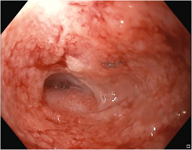
Endoscopic view of granulation tissue formation during endoscopic vacuum therapy exchange.
Read the full article online.
The information presented in Endoscopedia reflects the opinions of the authors and does not represent the position of the American Society for Gastrointestinal Endoscopy (ASGE). ASGE expressly disclaims any warranties or guarantees, expressed or implied, and is not liable for damages of any kind in connection with the material, information, or procedures set forth.

