
Post written by Ashutosh Mohapatra, MD, DM, and Sonmoon Mohapatra, MD, from Sai Institute of Gastroenterology and Liver Sciences, Odisha, India.

Our study describes a rare and educational case of early gastric signet ring cell carcinoma that presented as a solitary pinpoint gastric erosion during routine endoscopy. Despite the carcinoma’s deceptively benign appearance, careful endoscopic assessment, a high index of suspicion, and targeted biopsy led to the diagnosis of an early but biologically aggressive malignancy. This case highlights the importance of recognizing subtle endoscopic findings that may harbor significant pathology.

We report on this case because early gastric signet ring cell carcinoma often lacks typical endoscopic features and can be easily missed. In this patient, the lesion had been overlooked on a previous endoscopy and was detected at our center only because of meticulous examination and a high index of suspicion that an isolated gastric erosion could represent malignancy. This case shows that even a tiny solitary erosion may harbor carcinoma and underscores the importance of careful mucosal inspection, image-enhanced endoscopy, chromoendoscopy, and a low threshold for biopsy of subtle, unexplained lesions.
Single gastric erosions should not be overlooked, as they may represent early gastric cancer. Early signet ring cell carcinoma often appears as a subtle flat or slightly depressed pale lesion without obvious mucosal changes, making detection with white-light endoscopy challenging. Although magnifying narrow-band imaging improves diagnosis of early gastric cancer, its use is limited in undifferentiated carcinomas, which frequently spread beneath an intact surface epithelium.
In these cases, chromoendoscopy with indigo carmine can better delineate lesion borders. Therefore, biopsy of any unexplained pale, flat, depressed lesion or isolated gastric erosion remains essential for early diagnosis.
This case serves as a reminder that, in endoscopy, the smallest lesions may carry the greatest clinical significance. Maintaining vigilance, adhering to principles of high-quality endoscopic examination, and biopsying unexplained subtle mucosal abnormalities can facilitate earlier diagnosis of aggressive gastric cancers and potentially improve patient outcomes.
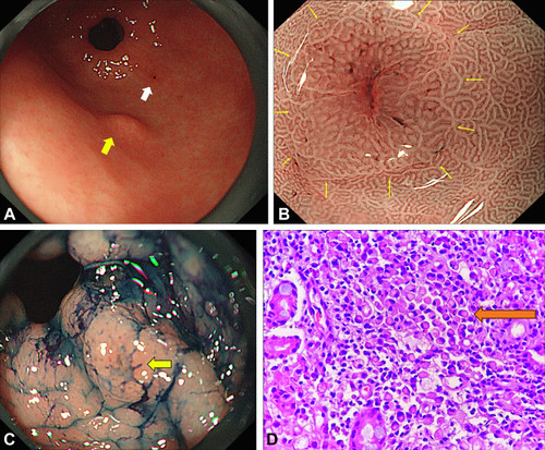
A, A 2-mm reddish erosion (Paris 0-IIc) was noted on the posterior wall of the prepyloric region (white arrow), approximately 2 cm distal to the subepithelial lesion (yellow arrow). B, Narrow-band imaging of the lesion showed marginal elevation with irregular or absent microsurface pattern at the center, without a clear demarcation line (yellow arrows). C, Chromoendoscopy showed a thorn-like encroachment, raising suspicion for malignancy (yellow arrow). D, Histopathologic examination with hematoxylin and eosin stain, original magnification ×400, showed signet ring cell carcinoma (orange arrow).
Visit iGIE’s Facebook, X/Twitter, LinkedIn, Instagram, and YouTube accounts for more content from the ASGE peer-reviewed journal that launched in December 2022 and has been PubMed indexed.
Read the full article online.
The information presented in Endoscopedia reflects the opinions of the authors and does not represent the position of the American Society for Gastrointestinal Endoscopy (ASGE). ASGE expressly disclaims any warranties or guarantees, expressed or implied, and is not liable for damages of any kind in connection with the material, information, or procedures set forth.

