
Post written by Tsukasa Ishida, MD, PhD, from the Department of Gastroenterology, Akashi Medical Center, Akashi, Japan.

We present the case of a 59-year-old woman with a giant pedunculated polyp in the transverse colon. The lesion was approximately 50 mm in diameter with an elongated stalk and considered technically difficult to resect safely by conventional snare EMR because of its size, mobility, and bleeding risk. Magnifying narrow-band imaging suggested an adenoma. We injected diluted epinephrine into the head and stalk of the polyp and placed 6 hemostatic clips near the stalk base.
Although immediate shrinkage was limited, repeat colonoscopy 3 weeks later showed marked reduction in the polyp head and stalk length. En bloc resection was then successfully performed using underwater EMR with a 25-mm snare. Histology confirmed a tubulovillous adenoma with negative margins, and the patient had no adverse events.
Giant pedunculated colorectal polyps can be challenging even for experienced endoscopists. In such cases, immediate resection may carry a substantial risk of bleeding or incomplete resection, but surgery may be more invasive than necessary for a benign lesion. This video demonstrates that delayed resection after epinephrine volume reduction and clip placement may be a useful strategy when immediate safe snaring is infeasible.
This case highlights the importance of adapting the treatment strategy to the morphology and technical risk of the lesion. When a giant pedunculated polyp cannot be safely captured with a snare at the initial procedure, epinephrine injection combined with clip placement may reduce blood flow and subsequently decrease lesion size. A short interval before repeat colonoscopy may convert a high-risk lesion into one that can be treated safely and completely by endoscopic resection.
To our knowledge, this case provides a useful example of interval volume reduction after epinephrine injection and clip placement for a giant pedunculated colorectal polyp. Careful optical diagnosis, patient selection, and close follow-up are essential when using this approach.
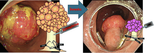
Endoscopic views before and 3 weeks after epinephrine injection and clip placement for a giant pedunculated polyp. A 50-mm polyp with a 50-mm stalk was treated with a 1:10,000 diluted epinephrine injection into the head and stalk, followed by the application of 6 clips at the stalk base. At the 3-week follow-up, the polyp had markedly decreased in size to approximately 20 mm.
Read the full article online.
The information presented in Endoscopedia reflects the opinions of the authors and does not represent the position of the American Society for Gastrointestinal Endoscopy (ASGE). ASGE expressly disclaims any warranties or guarantees, expressed or implied, and is not liable for damages of any kind in connection with the material, information, or procedures set forth.
