
Post written by Hussam Almasri, MD, from the from the University of North Dakota School of Medicine and Health Sciences, Fargo, North Dakota, and Rahul Karna, MD, Abubaker Abdalla, MBBS, Stuart K. Amateau, MD, PhD, and Martin Freeman, MD, from the Division of Gastroenterology, Hepatology, and Nutrition, University of Minnesota Medical Center, Minneapolis, Minnesota, USA.

We present a case of endoscopic management of malignant hilar biliary obstruction (Bismuth type IIIa cholangiocarcinoma) in a 74-year-old woman using dual-sector uncovered metal stent placement in a stent-in-stent (Y) configuration. After successful placement of a left hepatic stent, access to the right anterior sector was achieved with a guidewire.

However, standard devices, including a balloon catheter and stent delivery system, were unable to traverse the mesh of the first stent across a high-grade stricture. A spiral drill dilator (Tornus ES; Asahi Intecc Co, Aichi, Japan) was then used with controlled clockwise rotation to gently create a tract through the stent mesh and stricture. This enabled successful passage into the right anterior sector and placement of a second uncovered metal stent, completing the Y configuration and achieving effective dual-sector biliary drainage.

This case highlights how the spiral drill dilator can overcome one of the key technical challenges in hilar stenting, namely failure to traverse stent mesh in tight strictures. The video also demonstrates safety tips when using the dilator to minimize the risks of wire damage, stent deformation, or bile duct injury.
Endoscopists can learn that the Y configuration is feasible even in tight hilar strictures, although it can be technically challenging. Careful preprocedural planning and selection of appropriate tools are critical to success.
When standard devices fail to traverse the stent mesh, adjunctive tools such as a spiral drill dilator can be used to facilitate access. Importantly, these devices should be used with gentle, controlled movements to minimize the risk.
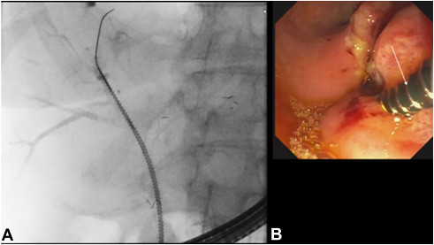
Fluoroscopic (A) and endoscopic images (B) showing a 7F spiral drill dilator (Tornus ES; Asahi Intecc Co, Aichi, Japan) (arrow) inserted over a 0.025-inch guidewire through the mesh of the left hepatic stent into the right anterior sector.
Read the full article online.
The information presented in Endoscopedia reflects the opinions of the authors and does not represent the position of the American Society for Gastrointestinal Endoscopy (ASGE). ASGE expressly disclaims any warranties or guarantees, expressed or implied, and is not liable for damages of any kind in connection with the material, information, or procedures set forth.
