
 Post written by Amrit K. Kamboj, MD, and Cadman L. Leggett, MD, from the Department of Internal Medicine and Division of Gastroenterology and Hepatology, Mayo Clinic, Rochester, Minnesota, USA.
Volumetric laser endomicroscopy (VLE) is an advanced imaging modality that uses optical coherence tomography to capture real-time, high-resolution, cross-sectional images of the esophagus. Epithelial glands are a known VLE feature associated with Barrett’s esophagus (BE) dysplasia. The focus of our study was to determine whether a systematic analysis of BE glands using VLE can determine the outcome of endoscopic resection.
Endoscopic surveillance is performed in patients with BE to diagnose and treat early neoplasia before it progresses to invasive cancer. Dysplastic BE lesions ≤ 2 cm in size can be targeted for en-bloc endoscopic mucosal resection (EMR). White light endoscopy may underestimate BE lesion size, leading to an incomplete resection. We felt that it was important to conduct this study to determine whether VLE can be used as a tool to guide endoscopic resection.
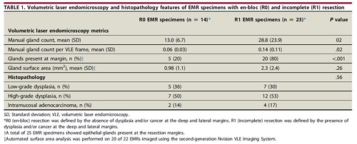
The study data set consisted of 37 EMR specimens from lesions with intent at endoscopic en-bloc resection that were imaged with VLE. Histologic en-bloc resection was achieved in 14 lesions and incomplete resection in 23 lesions. The en-bloc resection rate was 37.8%. A higher number of BE epithelial glands quantified with VLE was associated with incomplete resection, with a significant mean difference of 15.8 glands between groups (P=0.02). The presence of epithelial glands at the specimen margin was also associated with incomplete resection. We performed manual and automated analysis using a version of VLE image enhancement software that is now clinically available.
This is the first study to suggest that systematic quantification of a VLE feature can be used to determine the outcome of endoscopic resection. Epithelial gland quantification using VLE may be an important step in estimating BE lesion size and approach to endoscopic resection. Furthermore, computer-aided quantification tools may be applied to guide the endoscopist in estimating lesion size and resectability. The findings in this study need to be corroborated with in-vivo analysis of epithelial glands in order to establish the use of VLE in BE endoscopic therapy.
Our results suggest that VLE may have a potential role in providing guidance at the time of endoscopic resection. Piecemeal EMR or endoscopic submucosal dissection could be considered for a BE lesion imaged with VLE that is found to have a large epithelial gland burden. VLE with laser marking may then be used to outline the margins of the lesion by defining a region devoid of atypical epithelial glands.
Read the full article online.
The information presented in Endoscopedia reflects the opinions of the authors and does not represent the position of the American Society for Gastrointestinal Endoscopy (ASGE). ASGE expressly disclaims any warranties or guarantees, expressed or implied, and is not liable for damages of any kind in connection with the material, information, or procedures set forth.
