
Post written by Vivek Kumbhari, MD, from the Department of Medicine and Division of Gastroenterology and Hepatology, The Johns Hopkins Medical Institutions, Baltimore, Maryland, USA.
The success of gene-targeted therapy depends on 2 fundamental steps; the first being the intracellular delivery and the second is the genetic modification of the target cells. The first step can be carried out by either utilizing viruses to transduce the genes into the cells or other non-viral vectors. Although it has been proven to work, the use of viruses as a vector can have interference with the host immunity along with challenges in terms of accurate targeting of the relevant cell types and ensuring the durability of the genetic modification. For the above mentioned reasons, other forms of non-viral vectors with better safety profiles were developed to accomplish gene delivery; however, compared to viral vectors, their efficacy and durability is limited and further investigation is required for clinical adoption. Currently, what is needed is a gene-delivery method that combines selectivity, efficacy, safety, and durability in order to have more clinical advancement in the field of gene therapy.
Recently, a new method of gene delivery has gained popularity that does not involve the use of virus as a vector. The method is called Hydrodynamic Gene Delivery (HGD) due to the utilization of high pressure to inject genetic material through intra-vascular infusion. Interestingly, delivery to hepatocytes is particularly achievable at a higher levels compared to other organs presumably due to the extensive and porous circulation of blood within the liver at high surface area permitting the transmission of pressure. As a result, this method has been utilized to selectively target gene therapies in rodent and porcine livers to create or treat disease models. A successful macromolecule delivery to an ex-vivo human liver via intravascular hydrodynamic method was achieved as well. With this being said, carrying out (HGD) on in-vivo human subjects was not performed due to safety concerns. To achieve the gene delivery to the hepatocytes, it involves the infusion of a large volume under a high pressure likely resulting in lung and heart failure. A non-vascular hydrodynamic gene delivery would be the only viable method of materializing such a process in humans
We were able to adopt a novel non-vascular method of hydrodynamic gene delivery through the biliary route. Our hypothesis was that hydrodynamic injection of genetic material in a retrograde fashion through the bile duct during endoscopic retrograde cholangiopancreatograpjy (ERCP) procedure would overcome the major obstacles faced when targeting the liver in gene therapies. In our study, we used 12 pigs (40-50 kg) divided equally into 3 groups, and ERCP was performed by inflating a balloon catheter in the common hepatic duct thus creating a closed space while injecting a solution composed of plasmid/sleeping beauty (SB) mix. With this method, there was a closed space created between the point of injection and the liver parenchyma, allowing us to inject under pressure. Pigs were survived 21, 30, or 60 days. We were successful in transfecting a naked plasmid DNA with Sleeping Beauty Transposon to the hepatocytes. DNA-integration was achieved along with long-term expression of 3 oncogenic proteins (AKT, NICD, and beta-catenin), without having any hepatobiliary or cardiovascular adverse events.
After showing the feasibility of our extravascular method of hydrodynamic gene delivery, we are interested in directing our research towards determining the optimal application parameters for this method to achieve durable protein expression of our receptor genes in the hepatocytes of animal models.
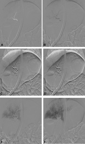
Figure 1. A-F, Fluoroscopic snapshot of swine bile duct distribution after contrast injection via ERCP. Rapid sequence fluoroscopic images (every 3 seconds) during injection of 30 mL of contrast medium at 2 mL/s. This resulted in acinarization of right and subsequently left liver segments without rupture of the bile duct wall (A-F). Therefore, these parameters were deemed optimal for hydrodynamic injection.
In summary, until now considerable progress has been achieved with highly efficacious techniques for gene therapy; however, the biggest obstacle is the absence of a safe and effective intracellular delivery method. As a solution, in our previous proof-of-concept study, we have shown that an extravascular method of hydrodynamic gene delivery is possible, safe, and effective. We believe our method is free of the well-known drawbacks of viral vectors as well as the potential adverse events of vascular HGD. It bears a significant potential for in-vivo and in-vitro intra-cellular transfer of genetic material. We are planning to pursue further research in this field, with the aim of developing this method and tackling specific disease models, and subsequently create a designated device.
Read the full article online.
The information presented in Endoscopedia reflects the opinions of the authors and does not represent the position of the American Society for Gastrointestinal Endoscopy (ASGE). ASGE expressly disclaims any warranties or guarantees, expressed or implied, and is not liable for damages of any kind in connection with the material, information, or procedures set forth.
