
Post written by Sho Suzuki, MD, PhD, from the Division of Gastroenterology and Hepatology, Department of Medicine, Nihon University School of Medicine, Tokyo, Japan.

We conducted this prospective, randomized, exploratory study to investigate the resection width and depth achieved by cold snare polypectomy (CSP) for small colorectal polyps compared to those achieved by hot snare polypectomy (HSP).
CSP was recently proposed as the primary technique for the resection of small colorectal polyps instead of HSP or endoscopic mucosal resection because of its low risk of adverse events. The resection width and depth achieved by polypectomy is associated with both curability and safety of polypectomy for colorectal tumors. However, the resection width and depth achieved by CSP compared to those achieved by HSP are still unknown.
This study showed that the mucosal defect size immediately after CSP was significantly larger than that after HSP, and it decreased within 1 day after CSP, while it increased for HSP. Additionally, the resection depth achieved by CSP was significantly more superficial than that achieved by HSP; CSP achieved resected submucosal tissue in only 24% cases, while HSP achieved resected submucosal tissue in over 80% of cases. However, both CSP and HSP successfully resected muscularis mucosa tissue in over 90% of cases in each group. These results suggest that CSP has sufficient curability for sub-centimeter colorectal polyps up to high-grade dysplasia, which is equivalent to HSP, and could account for a lower risk of adverse events such as bleeding and perforation after CSP compared to that after HSP. Thus, CSP is a reasonable treatment for sub-centimeter sized colorectal polyps.
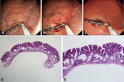
Figure 2. Endoscopic and histologic findings after cold snare polypectomy. A, A rectal polyp 9 mm in size (1 black or silver scale of measure device is 2 mm). B, Diameter of the mucosal defect immediately after cold snare polypectomy was 12 mm. C, Diameter of the mucosal defect 1 day later decreased to 8 mm. D, The resected specimen contained only muscularis mucosa tissue. E, The thickness of submucosal tissue was 0 μm.
Read the full article online.
The information presented in Endoscopedia reflects the opinions of the authors and does not represent the position of the American Society for Gastrointestinal Endoscopy (ASGE). ASGE expressly disclaims any warranties or guarantees, expressed or implied, and is not liable for damages of any kind in connection with the material, information, or procedures set forth.
