
Post written by Anne-Fré Swager from the Academic Medical Center, Amsterdam, the Netherlands

The focus of our study was to understand what we truly see on VLE: What do the VLE structures represent on histology? The aims were therefore to identify VLE features of Barrett’s neoplasia and to develop a VLE prediction score. We used a high-quality one-to-one correlated VLE-histology database.
Up to today, VLE features of Barrett’s neoplasia were mostly based on previous studies using older OCT systems and/or less precise correlation of OCT to histology. To fully understand VLE scans, it is important to precisely compare VLE images with the corresponding histology. With this high-quality VLE-histology database, we were able to further study and identify VLE features for Barrett’s neoplasia and develop these into a VLE prediction score.
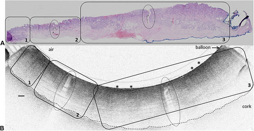
Figure 2. Histology-VLE match showing 2 ink markers (ovals) on histology (A) and VLE (B). The histology shows HGD (area 2) and EAC (area 3). On VLE, lack of layering is visible throughout the image, and increased VLE surface signal is also visible (area 3; asterisks). EAC, Early adenocarcinoma; ECM, electrocoagulation mark; HGD, high-grade dysplasia; VLE, Volumetric laser endomicroscopy. Scale bars represent 500 μm.
During 2 phases, 2 VLE experts scored in total 60 ex vivo VLE images blinded for the corresponding histology. Three VLE features independently predictive for BE neoplasia were identified and were developed into a prediction score: 1) lack of layering (6 points), 2) higher surface than subsurface signal (6 or 8 points for equal or higher surface signal), and 3) presence of irregular, dilated glands/ducts (5 points). The prediction score showed promising accuracy (AUC 0.81). A cut-off value of ≥ 8 points was associated with a sensitivity and specificity of 83% and 71%, respectively.
With an extensive supplementary section including many ex vivo VLE-histology correlations and descriptions of the VLE features of different mucosal types and focal features, this study provides a robust VLE atlas. Ongoing in vivo studies with larger sample sizes are warranted to validate the VLE neoplasia prediction score. Importantly, the VLE laser marking system has recently become available, which will enable in vivo correlation of VLE and histology for the first time.
Find the article abstract here.
The information presented in Endoscopedia reflects the opinions of the authors and does not represent the position of the American Society for Gastrointestinal Endoscopy (ASGE). ASGE expressly disclaims any warranties or guarantees, expressed or implied, and is not liable for damages of any kind in connection with the material, information, or procedures set forth.
