
Post written by Biswa Ranjan Patra, MD, DM, from Seth GS Medical College and KEM Hospital, Mumbai, India.

Removal of a deeply embedded esophageal self-expandable metal stent can be challenging, particularly after prolonged dwell time. Granulation tissue overgrowth may completely bury the stent, making retrieval difficult and increasing the risk of bleeding and perforation.
Although the stent-in-stent technique is commonly used, it may fail in selected cases. Specialized devices for powered endoscopic debridement offer an alternative, but their availability and cost may limit their use.
We describe a novel mechanical debridement technique using standard endoscopic accessories to remove a completely embedded, degenerated esophageal stent after conventional approaches had failed. This case involved an 18-year-old girl with a corrosive esophageal stricture and a stent that had remained in place for 26 months, despite an initial plan for removal after 6 weeks. Sequential stent-in-stent placement and repeated argon plasma coagulation (APC) failed to achieve adequate tissue necrosis and stent exposure.
This challenging situation prompted us to explore whether commonly available endoscopic accessories could provide an effective alternative to specialized equipment. The underlying principle was to combine controlled mechanical removal of overgrown tissue with the protective advantage of the embedded stent itself. The case also underscores the importance of structured follow-up and timely stent removal, particularly when stents are placed for benign esophageal disease.
Our technique combined circumferential APC with superficial mechanical debridement using open biopsy forceps, followed by deeper debridement using a distal attachment cap. The forceps were used to scrape ablated tissue, whereas the cap was applied tangentially to peel away residual granulation tissue. Debridement was performed in short segments, alternating with APC to help control mucosal oozing.
After 2 sessions, the stent was completely exposed and removed safely, along with residual wire fragments. No major adverse events occurred. Follow-up demonstrated mucosal healing, resolution of the stricture, and sustained symptom relief.
The key insight is that the embedded stent can serve as a protective scaffold, helping guide mechanical debridement while potentially reducing the risk of transmural injury. Combining superficial scraping with biopsy forceps and tangential peeling with a distal attachment cap allows progressive tissue removal using routinely available accessories.
This is a single-case experience, and the safety, reproducibility, and generalizability of the technique require further evaluation. We hope this article encourages endoscopists to think beyond conventional approaches when faced with difficult clinical scenarios. Sometimes, innovation lies not in developing a new device, but in discovering a new application for the tools already available. Meticulous technique, appropriate patient selection, and careful attention to safety remain essential.
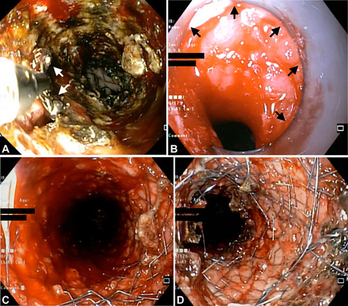
A, Endoscopic images showing superficial debridement of ablated granulation tissue with open biopsy forceps jaws (white arrows), (B) deeper debridement with distal attachment cap (black arrows), (C) partially exposed, and (D) completely exposed embedded stent.
Visit iGIE’s Facebook, X/Twitter, LinkedIn, Instagram, and YouTube accounts for more content from the ASGE peer-reviewed journal that launched in December 2022 and has been PubMed indexed.
Read the full article online.
The information presented in Endoscopedia reflects the opinions of the authors and does not represent the position of the American Society for Gastrointestinal Endoscopy (ASGE). ASGE expressly disclaims any warranties or guarantees, expressed or implied, and is not liable for damages of any kind in connection with the material, information, or procedures set forth.

