Post written by Dennis Wang, MD, from the Division of Gastroenterology and Hepatology, University of Rochester Medical Center Department of Medicine, Rochester, and Gregory Haber, MD, MASGE, from the Division of Gastroenterology and Hepatology, New York University Department of Medicine, New York, New York, USA.

We present a case of a patient with a symptomatic 4-cm esophageal leiomyoma, which was removed with submucosal tunneling endoscopic resection (STER). The lesion was known to be a leiomyoma, with no malignant potential, on previous EUS sampling, but in view of the obstructive symptoms, the patient strongly favored proceeding with STER.
After making a mucosotomy proximal to the lesion, we created a submucosal tunnel until the lesion was encountered rising into the submucosal space. The lesion was carefully dissected from its mucosal and submucosal attachments and resected including the muscularis propria attachments.

Recent advances in endoscopic devices and techniques have led to development of endoscopic methods for resecting subepithelial lesions of the GI tract that traditionally could only be removed with surgery. STER is particularly suitable for esophageal lesions because the relatively straight anatomy of the esophagus lends itself well to submucosal tunnel creation.
We demonstrate that STER is a feasible and safe method for managing such large esophageal lesions when performed at an institution that has experience with third space endoscopy. When compared with minimally invasive thoracoscopic surgery, STER has similar rates of en bloc resection and adverse events but shorter procedure time and decreased length of hospital stay.
There are many things for endoscopists to consider when performing STER. The initial mucosotomy should be at least 3 cm proximal to the lesion to allow stabilization of the endoscope in the tunnel for more accurate dissection. The mediastinum can be particularly treacherous in view of major vessels and lymphatics. We particularly prefer a coagulation-then-cut approach with the scissor-knife to avoid major bleeding.
As monopolar energy is used, we intentionally leave the most distal end of the lesion attached to conduct the current during piecemeal resection of the bulk of the lesion. A leiomyoma may not conduct current well, and the settings will need adjustment to cut through large pieces. Lesions larger than 3 cm can be difficult to remove en bloc, as they may become impacted at the mucosotomy site or the upper esophageal sphincter on extraction, so piecemeal resection of large lesions should be considered. This is particularly suitable for a leiomyoma with no malignant potential. This is the most common stromal tumor in the esophagus.
If the lesion straddles the cardia, we also have used a resection technique tunneling from above and below the lesion with clip closure at both ends. Endoscopists must be aware of the potential adverse events of STER, which include but are not limited to infection, bleeding, or pneumothorax. STER should only be performed on a symptomatic leiomyomata.
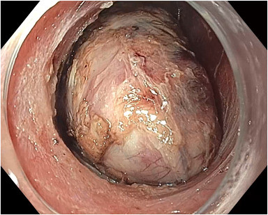
Leiomyoma dissected from the submucosal and muscular attachments.
Read the full article online.
The information presented in Endoscopedia reflects the opinions of the authors and does not represent the position of the American Society for Gastrointestinal Endoscopy (ASGE). ASGE expressly disclaims any warranties or guarantees, expressed or implied, and is not liable for damages of any kind in connection with the material, information, or procedures set forth.

