
Post written by Robert Bechara, MD, FRCPC, from the Division of Gastroenterology, Department of Medicine, Queen’s University, Kingston, Ontario, Canada, and Department of Gastroenterology, Cancer Institute Hospital, Japanese Foundation for Cancer Research, Tokyo, Japan.

Our review focuses on pharyngeal endoscopic submucosal dissection (ESD) for superficial pharyngeal squamous cell carcinoma. When these cancers are detected early and selected appropriately, pharyngeal ESD can offer a minimally invasive, organ-preserving treatment with excellent long-term disease-specific outcomes.
We summarize detection, optical diagnosis, indications, technical considerations, outcomes, adverse events, and practical steps for broader Western adoption. A central theme is that adoption will require more than resection skill alone: it requires systematic detection, focused training, appropriate equipment, airway planning, and close collaboration with otolaryngology and anesthesia.
This review was motivated by the clear gap between what has become achievable in high-volume Japanese pharyngeal ESD programs and what is currently available in most Western practice. In Japan, systematic pharyngeal examination and image-enhanced endoscopy have shifted detection toward earlier superficial lesions that may be amenable to endoscopic therapy. In contrast, early pharyngeal lesions in the West are often not deliberately sought or recognized, and pharyngeal ESD remains underutilized. We felt it was important to provide a practical framework for Western adoption, including training, patient selection, multidisciplinary collaboration, equipment, and follow-up, as early Canadian and Western experience continues to develop.
The outcomes reported in recent pharyngeal ESD series are encouraging. Across studies published from 2021 to 2025 and summarized in our review, en bloc resection was achieved in approximately 98.4% of cases, R0 resection in 78.5%, local recurrence occurred in 4.6%, lymph node metastasis in 4.8%, and 3- to 5-year disease-specific survival was 99.3%.
The most clinically important adverse event is laryngeal edema. Most cases are mild and managed conservatively, but approximately 2% require airway intervention. The review also emphasizes that good outcomes depend on careful patient selection: superficial disease without muscular invasion, nodal disease, or functionally excessive lesion extent. After resection, tumor thickness and high-risk histologic features help guide surveillance and the need for additional therapy.
The next steps for Western practice include education in structured pharyngeal examination and squamous optical diagnosis, focused training in pharyngeal ESD, and prospective Western registries to monitor outcomes and benchmark practice against high-volume Japanese centers.
We hope this article encourages endoscopists to pay deliberate attention to the pharynx during routine upper endoscopy and to develop collaborative pathways for patients with superficial pharyngeal carcinoma. Ultimately, pharyngeal ESD should be viewed as part of minimally invasive endoscopic oncology, but its success begins with detection.
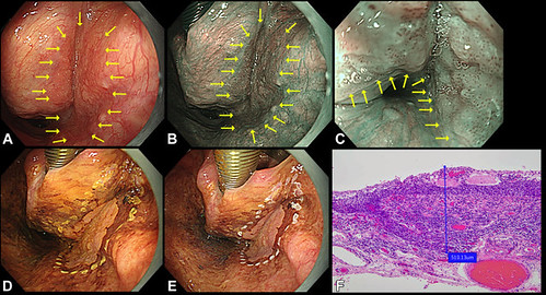
Superficial pharyngeal squamous cell carcinoma and measurement of subepithelial (SEP) squamous carcinoma. A and B, White-light and narrow-band imaging (NBI) views of a lesion in the right piriform sinus (yellow arrows delineate the lesion margins). C, Intrapapillary capillary loop changes are visible under NBI magnification (yellow arrows delineate the lesion margins). D and E, Lugol chromoendoscopy demonstrates the unstained area corresponding to the lesion. The pink color sign emerges after approximately 2 minutes. F, Histopathology (0.75% Lugol’s) demonstrating measurement of SEP tumor thickness from the epithelial surface to the deepest point of carcinoma invasion.
Read the full article online.
The information presented in Endoscopedia reflects the opinions of the authors and does not represent the position of the American Society for Gastrointestinal Endoscopy (ASGE). ASGE expressly disclaims any warranties or guarantees, expressed or implied, and is not liable for damages of any kind in connection with the material, information, or procedures set forth.

