
Post written by Serena Stigliano, MD, PhD, from the Therapeutic Endoscopy Department, Fondazione Policlinico Universitario Campus Bio-Medico, Rome, Italy.

Fluorescence confocal laser microscopy (FCM) allows imaging of tissues in the fresh state, with minimal preparation and without any damage, distortion, or loss of tissue.
We recently investigated its use in the evaluation of samples obtained with EUS fine-needle biopsy (EUS-FNB) in pancreatic solid lesions. We demonstrated a relevant performance in predicting the sample adequacy of EUS-FNB of pancreatic lesions (92.6%). The sensitivity of FCM was 100%, specificity 66.7%, accuracy 97%, positive predictive value 97%, and negative predictive value 100%. There was good agreement between the FCM diagnosis and the final histologic diagnosis with high diagnostic performances (Cohen’s κ coefficient, 0.95).
With this video, we aim to illustrate in detail the steps of this technique. The machine used is the microscope MAVIG VIVASCOPE 2500 by MAVIG GmbH (München, Germany). This video shows several important potential advantages of FCM with the microscope.
First, the chance to quickly analyze nonfixated, fresh tissue is an undisputed time-saving advantage. Second, this technique could add to the debate about using rapid onsite evaluation, reducing organizational issues and leading to significant cost-savings. Third, the real-time information about the adequacy of the sample could help with avoiding unnecessary needle passes, decreasing the time of the procedure and the risk of adverse events.
FCM also could offer a “distant diagnosis” by sharing the digital images for remote consulting with pathologists around the world and could give the opportunity of building a digital image library. Such a type of digital sharing also may be used to obtain a rapid pathology consultation in a hospital where a pathology unit is unavailable, allowing adequacy assessment by remote analysis.
The possibility of producing a digital image allows all the application tools of digital pathology, such as zooming in a high-power field, obtaining measurements of cellular and tissue structures, and saving the image for a re-evaluation.
In addition, the sample used for adequacy assessment undergoes formalin fixation and paraffin embedding that offers the chance of evaluating the same sample with the routine histologic procedures without losing it.
With this video, we want to show the ease and rapidity of the procedural steps of this new technique and, most of all, its interesting application in the rapid diagnosis of microhistological samples obtained with EUS-FNB.
In conclusion, this new technique can be successfully applied to cytological and histologic specimens. It provides fast information about the sample adequacy even in small specimens such as those obtained with EUS-FNB with good agreement with the final histologic report.
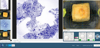 Fluorescence confocal laser microscopy software uses an algorithm to translate the acquired image information into colors that resemble hematoxylin and eosin.
Read the full article online.
The information presented in Endoscopedia reflects the opinions of the authors and does not represent the position of the American Society for Gastrointestinal Endoscopy (ASGE). ASGE expressly disclaims any warranties or guarantees, expressed or implied, and is not liable for damages of any kind in connection with the material, information, or procedures set forth.
