
Post written by Neal Mehta, MD, from the Department of Gastroenterology, Cleveland Clinic Foundation, Cleveland, Ohio, USA.

This video describes EUS-guided fine-needle biopsy (EUS-FNB) of an intraventricular mass in a 23-year-old woman.
The patient presented with unstable ventricular tachycardia requiring cardioversion and initiation of antiarrhythmic therapy. Cardiac MRI demonstrated a 7- x 7- x 3-cm mass, with vascular involvement concerning for malignancy, invading into the posterior left ventricular wall. Traditional transvascular cardiac biopsy methods were unfeasible because of the mass’s location, and cardiac surgery deemed the mass too large for resection.
Ultimately, the heart transplant team was consulted, but, given the lack of tissue diagnosis, transplant would be impractical if the mass were malignant.
After multidisciplinary discussion, EUS-FNB was planned. The procedure was performed in the cardiac intensive care unit with endotracheal intubation and deep sedation. In our video, we demonstrate that radial EUS was initially performed to delineate the cardiac anatomy and identify the mass.
Linear EUS was then performed for biopsy after the mass was identified. EUS from the gastroesophageal junction demonstrated approximately 3 cm of a heterogeneous, slightly hyperechoic mass within the left ventricle with no intervening vessels. Six passes with a 22-gauge SharkCore needle (Medtronic, Dublin, Ireland) were performed with on-site pathology present. The needle was directed through the esophageal wall, pericardium, and left ventricle and into the ventricular mass.
Postprocedurally, EUS-tip tamponade was performed without bleeding. No immediate or delayed adverse events occurred. Pathology revealed benign fibroadipose tissue. The patient ultimately underwent a successful cardiac transplant and is doing well.
As we know, EUS-FNB is a minimally invasive procedure commonly used for diagnostic purposes. Because of the accuracy and safety of EUS, both intraluminal and extraluminal lesions can be sampled.
However, few EUS-guided cardiac interventions have been published, as they are rarely performed because of potential risks. This video is important to demonstrate that the precise, real-time anatomic visualization of EUS-FNB makes it a safe, viable alternative for tissue acquisition when traditional sampling methods are problematic.
From this video case and technique described, we hope to impart that cardiac EUS-FNB may allow for safe and accurate diagnosis of cardiac masses in certain situations when traditional methods are unfeasible, tissue acquisition is necessary, and technical expertise is available. Because of the potential for adverse events and unique clinical scenarios that may require cardiac EUS-FNB, a multidisciplinary approach is highly recommended.
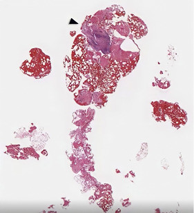 Pathology of the biopsy specimen demonstrating benign fibroadipose tissue (H&E, orig. mag. × 4).
Read the full article online.
The information presented in Endoscopedia reflects the opinions of the authors and does not represent the position of the American Society for Gastrointestinal Endoscopy (ASGE). ASGE expressly disclaims any warranties or guarantees, expressed or implied, and is not liable for damages of any kind in connection with the material, information, or procedures set forth.
