
Post written by Linda S. Yang, MBBS, from the Department of Gastroenterology, St Vincent’s Hospital Melbourne and the University of Melbourne, Melbourne, Australia.

At our tertiary referral center for Barrett’s esophagus, several endoscopic features have been observed in patients who were found to have buried Barrett’s mucosa on histology. This study evaluated the diagnostic accuracy of these endoscopic features and the frequency of endoscopically identifiable buried Barrett’s mucosa in patients with dysplastic Barrett’s esophagus.
Buried Barrett’s mucosa has been a clinical concern as it has been associated with dysplasia and neoplasia, especially in the post-treatment setting. However, there are no endoscopic features described in the literature, with conventional beliefs that endoscopes are unable to visualize subsquamous structures. There is a wide variation in endoscopic surveillance of the normal appearing neosquamous epithelium following endoscopic Barrett’s therapy with no established biopsy protocols. Therefore, buried Barrett’s mucosa and dysplasia may be missed in the absence of careful inspection for defined endoscopic features. We conducted this study with the aim of informing endoscopists that buried Barrett’s mucosa can be detected endoscopically with the trained eye.
The endoscopic features of buried Barrett’s mucosa were squamous epithelium, which is 1) darker pink on white light and darker brown on narrow-band imaging and/or 2) has a slightly raised or nodular appearance. It was also observed that either of these 2 features are frequently seen adjacent to a Barrett’s mucosa island.
In our cohort, buried Barrett’s mucosa was identified in 7%, including treatment-naïve patients. The proposed endoscopic features of buried Barrett’s were seen in 79% of patients with histology confirmed disease. An overlap between the endoscopic features of inflammation, reflux, and buried Barrett’s was observed.
This is the first study to describe endoscopic features of buried Barrett’s mucosa, which can predict the presence of buried Barrett’s mucosa and may also aid in detection of buried dysplasia or neoplasia. It has been thought that the majority of recurrences of intestinal metaplasia is endoscopically invisible and detected on random biopsies only. However, our results raise the question that perhaps “invisible” recurrences are in fact buried Barrett’s mucosa, which can be visible to the trained eye.
Future prospective studies are required to fine tune and validate the endoscopic criteria for identifying buried Barrett’s mucosa.
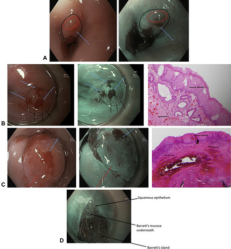
Figure 1. Proposed endoscopic features of buried Barrett’s mucosa. A and B, Darker pink (white-light endoscopy) or dark brown (narrow-band imaging) area in relation to surrounding squamous epithelium. In a patient after previous endoscopic therapy for dysplastic Barrett’s mucosa, surveillance endoscopy with high-definition white light shows the outlined area (in black) with darker pink mucosa (red circle). This is adjacent to an island of Barrett’s mucosa (arrow). The same area on narrow-band imaging shows the darker brown mucosa (red circle) adjacent to a Barrett’s island (arrow). This suggests subsquamous extension of Barrett’s island and highlights the importance of assessing islands of Barrett’s and the surrounding mucosa in the post-treatment setting. A, Within this ring of slightly elevated area (black outline), there is darker pink mucosa (black arrows) covered by squamous epithelium, with adjacent islands of Barrett’s mucosa (blue arrows). Narrow-band imaging demonstrates corresponding dark brown area (black arrows), containing the buried Barrett’s mucosa. Histologic examination demonstrates low-grade dysplasia within the buried Barrett’s mucosa underneath a clear layer of squamous epithelium. C, Buried Barrett’s mucosa associated with an island of Barrett’s mucosa. The endoscopic photos demonstrate neosquamous epithelium (red arrow) adjacent to an island of Barrett’s mucosa (blue arrow). Histologic examination of the endoscopic mucosal resection specimen demonstrates the Barrett’s island and a subsquamous extension of Barrett’s mucosa containing high-grade dysplasia. D, Buried Barrett’s mucosa seen in continuation with an island of Barrett’s mucosa. On narrow-band imaging, pearlescent darker brown squamous epithelium is seen (arrow), overlying Barrett’s mucosa underneath. This buried Barrett’s mucosa is in continuation with a Barrett’s island.
Read the full article online.
The information presented in Endoscopedia reflects the opinions of the authors and does not represent the position of the American Society for Gastrointestinal Endoscopy (ASGE). ASGE expressly disclaims any warranties or guarantees, expressed or implied, and is not liable for damages of any kind in connection with the material, information, or procedures set forth.
