
Post written by Tae Hoon Lee, MD, PhD, and Do Hyun Park, MD, PhD, from the Department of Internal Medicine, Soonchunhyang University College of Medicine, Cheonan Hospital, Cheonan, and the Department of Internal Medicine, University of Ulsan College of Medicine, Asan Medical Center, Seoul, South Korea.

Membrane-covered self-expandable metal stents (SEMS) have been developed to prolong patency of stents by reducing tissue hyperplasia or tumor ingrowth. However, efficacy of SEMS is attenuated by stent clogging due to biofilm formation on the inner covering surface of membrane. Therefore, we tried to evaluate the efficacy and safety of SEMS covered with a silicone membrane containing integrated silver particles (Ag-P) to prevent biofilm formation in malignant distal biliary obstruction.
Anti-biofilm- and anti-inflammation-coated stent membranes may prolong stent patency and are expected to help improve both patients’ quality of life and the cost-effectiveness of treatment by avoiding additional unnecessary re-intervention procedures related to stent dysfunction.
Duodeno-biliary reflux can result in bacterial deposits and biofilm formation on the inner coating surface membrane of SEMS, and result in sludge or stone formation. Thus, functional improvement of covered SEMS is required to prolong stent patency and survival by improving stent function.
SEMS covered with a silicone membrane containing integrated Ag-P resulted in promising stent patency without serious stent or Ag-P-related adverse events. This type of integrated-Ag-P membrane may reduce sludge impaction related to biofilm formation, which may prolong stent patency.
However, further large-scale randomized comparative studies comparing this new stent with conventional covered metal stents may be warranted to confirm the ability of the SEMS covered with a silicone membrane containing integrated Ag-P to inhibit biofilm and sludge formation.
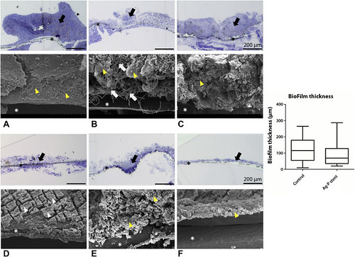
Figure 4. Light microscopy images and the corresponding scanning electron micrographs of biofilm on luminal surfaces of stents (A-F panels, longitudinal column). Details of biofilm formed on the inner surface of 6 different stents observed by light microscopy and SEM. Cross-sectional view of the silicone membrane (asterisk) and biofilm (black arrow) from 3 stents in the control group (A-C; patient numbers 4, 6, 12) and 3 stents in the Ag-particle-integrated test group (D-F; patient numbers 9, 10, 13) were observed by light microscope (orig. mag., ×50). Scale bar, 200 μm. The average biofilm thickness measured from 18 stents in the control group (n = 18) and 7 stents in the Ag-particle-integrated Ag-P stent group (n = 7) is shown in the bar graph bar. Rich dietary fibers, amorphous material, and bile salts are indicated by white arrows, yellow arrowheads, and white arrowheads, respectively. SEM, Scanning electron microscopy; Ag-P, silver nanoparticles.
Read the full article online.
The information presented in Endoscopedia reflects the opinions of the authors and does not represent the position of the American Society for Gastrointestinal Endoscopy (ASGE). ASGE expressly disclaims any warranties or guarantees, expressed or implied, and is not liable for damages of any kind in connection with the material, information, or procedures set forth.
