

 Post written by Tae Hoon Lee, MD, PhD, and Jong Ho Moon, MD, PhD, from the Department of Internal Medicine, SoonChunHyang University College of Medicine, Cheonan, Republic of Korea.
Although the efficacy of palliative bilateral biliary drainage using self-expandable metal stents (SEMS) has been demonstrated, it is unclear which bilateral deployment method using SEMS is optimal for advanced malignant hilar biliary strictures (MHS). Therefore, this pilot study compared bilateral stent-in-stent (SIS) with stent-by-stent (SBS) deployment for advanced MHS; we tried to compare outcomes such as adverse events, technical and clinical success, re-intervention, therapeutic outcomes, stent patency, and survival duration.
The methods for achieving adequate stent patency and avoiding adverse events when deploying multiple or bilateral SEMS for an advanced MHS remain controversial. Also, there are no well-designed comparative results. This is the first multicenter comparative study of bilateral SIS and SBS methods to prove the efficacy of bilateral stenting methods using SEMS.
Bilateral SIS and SBS deployment might show similar efficacy in terms of total adverse events, technical and clinical success rates, and revision efficacy. Technically, the SIS method was not more difficult than SBS for experts. The stent patency rate was not significantly different between the 2groups, but it tended to be higher at both 3 and 6 months in the SIS group without a statistical difference. Based on this comparative study, we think that both SIS and SBS methods are technically feasible and have similar efficacy even in high-grade MHS.
Although there was no statistical difference between the 2 methods, SIS deployment might have a tendency toward a higher rate of stent patency without a significant difference. The SIS method may be more physiologically sound than the SBS method. Further large-sized, long-term studies involving multiple centers are required to validate our pilot results.
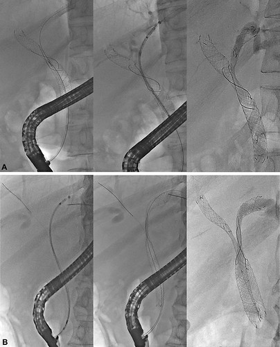
Figure 2. A, Stent-in-stent (SIS) deployment. Y-shaped configuration of SIS showing the second stent passing through the first deployed metal stent. First stent was deployed in the right intrahepatic duct (IHD) (left), second stent through the wire mesh of first stent (middle), and final configuration of Y-shaped SIS deployment (right). B, Stent-by-stent (SBS) deployment. Parallel configuration of the SBS positioned above the level of the papilla with the same distal margin. First stent was inserted in left IHD (left) and deployed (middle). After deployment of the second stent in the right IHD, SBS deployment was completed with same distal margin of stents (right).
Read the full article online.
The information presented in Endoscopedia reflects the opinions of the authors and does not represent the position of the American Society for Gastrointestinal Endoscopy (ASGE). ASGE expressly disclaims any warranties or guarantees, expressed or implied, and is not liable for damages of any kind in connection with the material, information, or procedures set forth.
