
Post written by JianYing Bai, PhD, from the Department of Gastroenterology, Xinqiao Hospital, Third Military Medical University, Chongqing, China.
We focused on the effectiveness and safety of endoscopic transplantation of autologous esophageal mucosa in preventing stricture formation after circumferential ESD.
Esophageal stricture is a very common adverse event after ESD, especially with resection of nearly circumferential lesions or whole-circumferential lesions with a rate of 88% to 100%. Dilation is the most common treatment of esophageal strictures and always with long therapy duration. Several means have been attempted to prevent post-ESD stricture with varying effects.
Our results showed that transplantation of autologous esophagus mucosa relieved the severity of esophageal stricture after circumferential ESD. This method also reduced the time of dilation after esophageal stricture. In this context, our study provided a novel alternative method to reduce the stricture formation after esophageal circumferential ESD. Long-term outcomes should be followed. There is still long way to go to efficiently fight esophageal cancer and related post-ESD strictures.
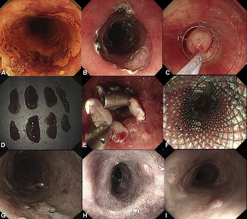
Figure 1. Procedure of endoscopic esophageal mucosa transplantation. A, 1.5% Lugol’s solution to further determine the extent of the lesion. B, Resection of the invaded mucosa involved with the entire circumference of the esophageal lumen by using endoscopic submucosal dissection. C, Autologous esophageal mucosal tissue was excised by EMR by using a crescent snare. D, Normal mucosa was cut into 3-mm by 10-mm pieces. E, Mucosal patches were attached to the ulcer surface by using clips. F, A covered metal mesh stent was used to make the mucosa patches fully contacted and firmly adhere to the ulcer surface. G-I, One case undergoing circumferential endoscopic submucosal dissection with esophageal stricture prevented by the application of the transplantation of esophageal mucosa (case 8). G, One week after transplantation, epithelialization was observed at the center of the ulcerative site. H, Three weeks after transplantation, complete epithelization was observed. I, Twenty-four weeks after transplantation, no evidence of stricture was observed.
Read the full article online.
The information presented in Endoscopedia reflects the opinions of the authors and does not represent the position of the American Society for Gastrointestinal Endoscopy (ASGE). ASGE expressly disclaims any warranties or guarantees, expressed or implied, and is not liable for damages of any kind in connection with the material, information, or procedures set forth.
