
Post written by Gian Eugenio Tontini, MD, PhD, and Helmut Neumann, MD, PhD, from the Gastroenterology and Digestive Endoscopy Unit, IRCCS Policlinico San Donato, San Donato Milanese, Milan, Italy, and the Department of Interdisciplinary Endoscopy, I. Medizinische Klinik und Poliklinik, University Hospital, Mainz, Germany.
Our study focuses on the value of Confocal Laser Endomicroscopy (CLE) for the characterization of mucosal inflammatory and architectural changes in the colon-rectum of Crohn’s disease (CD) patients, thereby predicting long-term clinical outcomes.
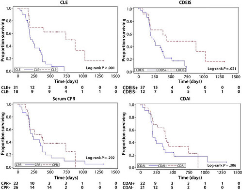
Supplementary Figure 1. Kaplan-Meier curves for the estimated 4-year survival rate of medical treatment escalation according to baseline confocal laser endomicroscopy (CLE), Crohn’s disease endoscopic index of severity (CDEIS), C-reactive protein (CRP), and Crohn’s disease activity index (CDAI).
The assessment of reliable prognostic factors can be crucial for early intervention and treat-to-target strategies in CD. Both endoscopic and microscopic healing from mucosal inflammation have been shown to be associated with improved IBD outcomes. Two recent pilot studies suggested that confocal findings from the terminal ileum of IBD patients correlate with the risk of future clinical relapse, hospitalization, or surgery. In contrast, our study focused on the in vivo characterization of microscopic architectural (ie, crypt architecture abnormality) and inflammatory (ie, focal cryptitis) changes in the large bowel, which are regarded as hallmarks for histopathological evaluation in active CD.
CLE enables in vivo characterization of microscopic tissue features of CD mucosal changes conventionally used by standard histopathology to confirm diagnosis and assess disease activity. The combined presence of focal cryptitis and discontinuous crypt architectural abnormality appears as a promising predictor of important clinical outcomes, such as treatment escalation and transmural adverse events. In this context, confocal findings were superior to CDEIS, CRP, and CDAI assessment. The potential of CLE was substantial in the short term but disappeared after 1-year follow-up. Thus, CLE assessment of mucosal signs of CD activity may allow for early risk stratification related to strong clinical outcomes, thereby potentially refining the timing of treatment strategies targeting mucosal inflammation in CD.
To the best of our knowledge, no previous study has never assessed the predictive value of confocal imaging over the impact of standard clinical and endoscopic disease-course indicators in the field of IBD. Therefore, the present findings represent a unique picture and the leading reference for further case-control studies or multivariate analyses on this topic.
Read the full article online.
The information presented in Endoscopedia reflects the opinions of the authors and does not represent the position of the American Society for Gastrointestinal Endoscopy (ASGE). ASGE expressly disclaims any warranties or guarantees, expressed or implied, and is not liable for damages of any kind in connection with the material, information, or procedures set forth.
