
Post written by Yoshihiro Furuichi, MD, PhD, from the Department of Gastroenterology and Hepatology, Tokyo Medical University, Tokyo, Japan.

Dual red imaging (DRI) is a novel image-enhanced endoscopy technique that can increase the visibility and predict the depth of esophageal varices (EVs). The recurrence rate of EVs after endoscopic-injection sclerotherapy (EIS) reportedly decreases by intravariceal injection of a sclerosant. We evaluated prospectively whether the EIS intra-success rate was increased by DRI compared to the white-light imaging (WLI) mode. As a result, DRI increased the EIS intra-success rate and decreased the recurrence rate of EVs compared to WLI.
Treatment of high-risk EVs is thought to be necessary for preventing massive bleeding from EVs, which often prove to be fatal. However, the chance of liver transplantation is much lower in Japan than in Western European countries because of donor problems. Therefore, the method of complete removal of EVs is desired. The recurrence rate of EVs after EIS using intravariceal sclerotherapy injection method is much lower than that after endoscopic variceal ligation, but the intra-injection success rate is low in community hospitals because the technique is difficult.
Our present prospective study demonstrated that DRI can increase the success rate of intravariceal injection on the first puncture and the total punctures in EIS, and it can result in a lower recurrence rate and a shorter operating time.
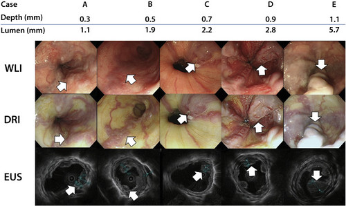
Figure 3. Endoscopic and EUS images. A, to E, The white light, dual red imaging, and EUS images of the esophageal varices are shown in the order of the depth from the epidermis. A, The shallowest depth of esophageal varices. E, The deepest depth of esophageal varices. The esophageal varices with a shallower depth became more clearly visualized by dual red imaging. The arrows show the particular esophageal varices. WLI, white-light imaging; DRI, dual red imaging.
We think the high success rate of intra injection in DRI group is related to the length of the needle used in performing EIS. In the sub-analysis to identify the cause, the Pearson correlation coefficient between the needle length and the depth plus luminal diameter in the success group was higher than that in the failure group. This indicates that the appropriate adjustment of the needle length suited to the depth and luminal diameter of EVs increases the success rate of EIS and that DRI can help achieve this by predicting the depth of EVs. We think that the higher success rate of intravariceal injection enabled us to deliver effective treatment with fewer EIS punctures (shorter operating time) and a lower recurrence rate.
For detailed examination of the depth and luminal diameter of EVs, EUS is very useful. However, many institutions do not have an EUS device for economic reasons. DRI can solve this problem. The DRI function will be installed in the next endoscopic light source device to be released. If DRI can be used in many community hospitals, the success rate of EIS and the operation time certainly will be improved by prediction of the depth of EVs.
Read the full article online.
The information presented in Endoscopedia reflects the opinions of the authors and does not represent the position of the American Society for Gastrointestinal Endoscopy (ASGE). ASGE expressly disclaims any warranties or guarantees, expressed or implied, and is not liable for damages of any kind in connection with the material, information, or procedures set forth.
