
Post written by Toyoki Kudo, MD, PhD, from the Digestive Disease Center, Showa University Northern Yokohama Hospital, Yokohama, Japan.

Endoscopic diagnosis makes it possible to differentiate between colorectal carcinomas with slight submucosal invasion (SM-s), defined as carcinomas with a vertical invasion depth of less than 1000 µm in the submucosal layer, and massive submucosal invasion (SM-m), defined as those with an invasion depth greater than 1000 µm. This distinction is important for planning subsequent treatment strategies because lesions up to SM-s have almost no risk of lymph node metastasis, whereas those with SM-m have a 10–15% risk of lymph node metastasis.
However, one of the problems with the currently used EC classification is its inability to differentiate between SM-s and SM-m. If EC3a findings were to be considered a good index for lesions up to SM-s, this would make it difficult to differentiate between SM-s and SM-m because considerably more SM-m would be diagnosed with EC3a. Therefore, we believe that there is a need to reconsider EC3a findings that can differentiate between SM-s and SM-m.
In this article, we determined if factors related to EC findings could be used to create a new SM-m index to improve the diagnostic accuracy of EC3a. We also retrospectively compared the diagnostic performance of this new EC classification with that of NBI and pit-pattern for evaluating invasion depth. We analyzed 8 aspects of EC images of colorectal tumors to determine if they were associated with SM-m or worse. Multivariate analysis indicated that unclear glandular lumens (ULs), high degree of nuclear enlargement (HNE), and multilayered nuclei (MNs) were the most useful factors for making a diagnosis of SM-m or worse. Based on these results, we divided tumors in the EC3a category into low-grade or high-grade and investigated the diagnostic performance of this sub-classification. This distinction was associated with a sensitivity of 88.9%, specificity of 91.3%, positive predictive value of 75.0%, negative predictive value of 96.6%, accuracy of 90.8%, and a positive likelihood ratio of 10:2. Furthermore, the sensitivity, negative predictive value, and accuracy of this new EC classification were significantly higher than those of NBI and pit-pattern classification.
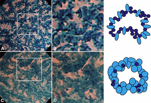
Figure 5. EC3a subclassification. A and B, EC3a low grade, characterized by numerous oval strongly stained nuclei, low degree of enlarged nuclei, low degree of multilayering, and comparatively sharp nuclear morphology. C and D, EC3a high grade, characterized by numerous irregularly shaped nuclei, high degree of enlarged nuclei, high degree of multilayering, and comparatively dull nuclear morphology. EC, endocytoscopy.
Based on our findings, we concluded that EC findings of ULs, HNE, and MNs are important risk factors for SM-m or worse. Furthermore, our EC3a sub-classification taking these findings into consideration could improve the diagnosis of SM-m or worse in patients with colorectal neoplasms.
Find the article abstract here.
The information presented in Endoscopedia reflects the opinions of the authors and does not represent the position of the American Society for Gastrointestinal Endoscopy (ASGE). ASGE expressly disclaims any warranties or guarantees, expressed or implied, and is not liable for damages of any kind in connection with the material, information, or procedures set forth.

Good Information. Thanks for sharing.