
Post written by Riku Nakajima, MD, Wataru Sano, MD, Sachiko Shimizu, MD, Naoki Sugimura, MD, PhD, Daizen Hirata, MD, PhD, Mineo Iwatate, MD, PhD, Mikio Fujita, MD, PhD, and Yasushi Sano, MD, PhD, from the Gastrointestinal Center, Sano Hospital, Kobe, Japan.

Our article presents 4 patients who underwent underwater endoscopic submucosal dissection for large colonic lipomas. These lesions can be difficult to resect because their size makes it difficult to see the base.
After we filled the lumen with saline, the lipoma floated upward. This provided continuous countertraction and exposed the base and dissection plane. The video shows how we used this effect while dissecting beneath the lipoma.
The benefit of saline immersion is much easier to understand when seen than when described in words. As the lipoma floats, its base becomes clearer, and the working space changes. We wanted to share this simple observation because it helped us address a common difficulty in the endoscopic treatment of large colonic lipomas: maintaining a clear view of where to dissect.
Buoyancy can act as a source of traction during underwater endoscopic submucosal dissection for a colonic lipoma. Rather than relying only on an additional traction device, the endoscopist may be able to use the lesion’s own physical properties to expose the dissection plane. Careful identification of that plane remains essential, and bleeding can quickly obscure the view under saline. Our 4 cases feature a useful technical option for selected lesions; a larger study would be needed to compare it with other resection methods.
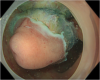
Endoscopic images of underwater endoscopic submucosal dissection for large colonic lipomas (case 3). Because of the low specific gravity of adipose tissue, the lipoma is floating just beneath the surface of the normal saline solution. A distinct reflection of the specimen is visible on the saline surface (top), highlighting the buoyancy characteristic of colonic lipomas.
Read the full article online.
The information presented in Endoscopedia reflects the opinions of the authors and does not represent the position of the American Society for Gastrointestinal Endoscopy (ASGE). ASGE expressly disclaims any warranties or guarantees, expressed or implied, and is not liable for damages of any kind in connection with the material, information, or procedures set forth.

