
Post written by Kambiz Kadkhodayan, MD, and Muhammad K. Hasan, MD, FACG, FRCP (Glasg), from the Center for Interventional Endoscopy, AdventHealth Orlando, Orlando, Florida, USA.

We present a patient with obstructive jaundice and a flat (type 1) ampulla in whom conventional biliary cannulation using a standard sphincterotome was unsuccessful despite prolonged attempts. Given the unfavorable papillary morphology and need for biliary access, we proceeded with needle-knife sphincterotomy.
Prior to initiation of the incision, a targeted submucosal injection of dyed lifting solution was placed just above the ampullary orifice. This created a controlled submucosal cushion and enhanced visual differentiation among mucosa, submucosa, and deeper layers during dissection. Layer-by-layer needle-knife sphincterotomy was then performed, resulting in successful identification and cannulation of the bile duct without adverse events.

Needle-knife sphincterotomy continues to be an essential rescue technique, yet many endoscopists approach it with caution because of concerns regarding bleeding, perforation, and poor visualization, particularly in flat ampullae with nondilated ducts. This case highlights a simple, readily available adjunct that may improve anatomic orientation during freehand dissection. Our intent was to demonstrate a pragmatic technique that can make an inherently high-risk maneuver more controlled and predictable, especially for operators who do not perform needle-knife access frequently.
This video emphasizes the importance of tissue plane identification during advanced cannulation. Submucosal dye injection can serve as a visual guide, helping the endoscopist recognize when dissection remains within the submucosal space versus when it drifts toward deeper, higher-risk layers. This approach may be particularly useful in patients with unfavorable papillary anatomy, where conventional landmarks are less distinct.
This technique should be viewed as an adjunct to established principles of advanced biliary access. To our knowledge, this represents the first video description of submucosal injection used specifically to facilitate needle-knife sphincterotomy, and further experience will be needed to define its role.
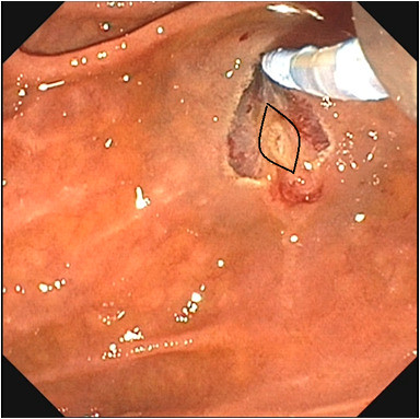
Endoscopic image outlining the ampullary os (black lines). Note its distinct appearance (does not stain with methylene blue) when compared with adjacent submucosal tissue (stains with methylene blue).
Read the full article online.
The information presented in Endoscopedia reflects the opinions of the authors and does not represent the position of the American Society for Gastrointestinal Endoscopy (ASGE). ASGE expressly disclaims any warranties or guarantees, expressed or implied, and is not liable for damages of any kind in connection with the material, information, or procedures set forth.
