
Post written by Naoya Tada, MD, PhD, Akira Dobashi, MD, PhD, and Kazuki Sumiyama, MD, PhD, from the Department of Endoscopy, The Jikei University School of Medicine, Tokyo, Japan.

Our video demonstrates a combined technique of scar incision and endoscopic balloon dilation (EBD) for severe benign esophageal strictures following endoscopic submucosal dissection or esophagectomy. After identifying areas of severe fibrosis, we selectively create one or more linear mucosubmucosal incisions using a needle-knife.
These incisions are placed specifically at sites where fibrotic tension is most pronounced or where conventional EBD has repeatedly failed to induce adequate tearing. Once sufficient targeted tension is released by scar incision, EBD is performed, allowing expansion of the entire stricture segment with improved safety and efficacy.
Conventional EBD does not allow control over where a tear occurs. Tears often arise randomly or recur in the same weaker area, leaving the most fibrotic regions undertreated. In other cases, no tear occurs at all, preventing adequate reduction of fibrotic tension. These limitations of conventional EBD can lead to insufficient dilation, repeated procedures, and occasionally perforation.
By placing incisions precisely at sites where tension is highest or needs to be reduced, we can intentionally guide where subsequent dilation forces are applied. This allows more predictable tension release, minimizes uncontrolled tearing, and may reduce the number of EBD sessions required.
We felt it was important to present these cases because they directly address the fundamental limitation of conventional dilation: the inability to control fibrotic tension. To our knowledge, this technique offers a practical and reproducible strategy to optimize outcomes in highly fibrotic strictures.
The 5 cases in this video each highlight different situations in which scar incision adds value. We hope that endoscopists encountering similar patterns will consider adopting EBD with scar incision. Precise tension control might offer a meaningful approach to reducing the number of dilation sessions and improving patients’ symptoms. The technique is conceptually simple and can be incorporated into clinical practice.
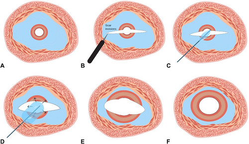
The schematic image of the endoscopic balloon dilation (EBD) with scar incision. A, Benign esophageal stricture. B, Scar incisions. C, Inserting the balloon. D, Balloon dilation. E, After the EBD with scar incision. F, Achieving the resolution of the stricture.
Read the full article online.
The information presented in Endoscopedia reflects the opinions of the authors and does not represent the position of the American Society for Gastrointestinal Endoscopy (ASGE). ASGE expressly disclaims any warranties or guarantees, expressed or implied, and is not liable for damages of any kind in connection with the material, information, or procedures set forth.
